Mycobacterium smegmatis
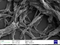
Mycobacterium smegmatis is an acid-fast bacterial species in the phylum Actinomycetota and the genus Mycobacterium. It is 3.0 to 5.0 µm long with a bacillus shape and can be stained by Ziehl–Neelsen method and the auramine-rhodamine fluorescent method. It was first reported in November 1884 by Lustgarten, who found a bacillus with the staining appearance of tubercle bacilli in syphilitic chancres. Subsequent to this, Alvarez and Tavel found organisms similar to that described by Lustgarten also in normal genital secretions (smegma). This organism was later named M. smegmatis.[1]
| Mycobacterium smegmatis | |
|---|---|
 | |
| Scientific classification | |
| Domain: | Bacteria |
| Phylum: | |
| Class: | |
| Order: | |
| Family: | |
| Genus: | |
| Species: | M. smegmatis |
| Binomial name | |
| Mycobacterium smegmatis (Trevisan 1889) Lehmann & Neumann 1899 | |
Some species of the genus Mycobacterium have recently been renamed to Mycolicibacterium, so that M. smegmatis is now Mycolicibacterium smegmatis.[2][3]
Virulence
M. smegmatis is generally considered a non-pathogenic microorganism; however, in some very rare cases, it may cause disease.[4]
Use in research
M. smegmatis is useful for the research analysis of other Mycobacteria species in laboratory experiments. M. smegmatis is commonly used in work on the Mycobacterium genus due to it being a "fast grower" and non-pathogenic. M. smegmatis is a simple model that is easy to work with, i.e., with a fast doubling time and only requires a biosafety level 1 laboratory. The time and heavy infrastructure needed to work with pathogenic species prompted researchers to use M. smegmatis as a model for mycobacterial species.
M. smegmatis shares the same peculiar cell wall structure of M. tuberculosis and other mycobacterial species.[5] It is also capable of oxidizing carbon monoxide aerobically, as is M. tuberculosis.
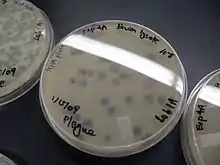
M. smegmatis is readily cultivatable in most synthetic or complex laboratory media, where it can form visible colonies in 3–5 days. These properties make it a very attractive model organism for M. tuberculosis and other mycobacterial pathogens. M. smegmatis mc2155 is also used for the cultivation of mycobacteriophage.
Genetics and genomics
The genomes of multiple strains of M. smegmatis have been sequenced by TIGR and other laboratories, including the "wild-type" (mc2 155) and some antibiotic-resistant strains (4XR1/R2).[6] The genome of strain mc2155 is ~6,9 Mbp long and encodes ~6400 proteins[7] which is relatively large for bacteria (for comparison, the genome of E. coli encodes about 4000 proteins).
This species shares more than 2000 homologous genes with M. tuberculosis and thus is a good model organism to study mycobacteria in general and the highly pathogenic M. tuberculosis in particular.
The discovery of plasmids, phages, and mobile genetic elements has enabled the construction of dedicated gene-inactivation and gene reporter systems. The M. smegmatis mc2155 strain is hypertransformable, and is now the work-horse of mycobacterial genetics.
Transformation
Transformation is a process by which a bacterial cell takes up DNA that had been released by another cell into the surrounding medium, and then incorporates that DNA into its own genome by homologous recombination (see Transformation (genetics)). Strains of M. smegmatis that have particularly efficient DNA repair machinery, as indicated by their greater resistance to the DNA damaging effects of agents such as UV and mitomycin C, proved to be the most capable of undergoing transformation.[8] This suggests that transformation in M. smegmatis is a DNA repair process, presumably a recombinational repair process, as it is in other bacterial species.[9]
Conjugation
Conjugal DNA transfer in M. smegmatis requires stable and extended contact between a donor and a recipient strain, is DNase resistant, and the transferred DNA is incorporated into the recipient’s chromosome by homologous recombination. However, in contrast to the well-known E. coli Hfr conjugation system, in M. smegmatis all regions of the chromosome are transferred with comparable efficiencies and mycobacterial conjugation is chromosome, rather than plasmid based. Gray et al.[10] reported substantial blending of the parental genomes resulting from conjugation and referred to this blending as reminiscent of that seen in the meiotic products of sexual reproduction (see Origin of sexual reproduction).
DNA repair
M. smegmatis relies on DNA repair pathways to resist DNA damage. Double-strand breaks are especially threatening to bacterial viability. M. smegmatis has three options for repairing double-strand breaks; homologous recombination (HR), non-homologous end joining (NHEJ), and single-strand annealing (SSA).[11] The HR pathway of M. smegmatis is the major determinant of resistance to ionizing radiation and oxidative DNA damage. This pathway involves exchange of information between a damaged chromosome and another homologous chromosome in the same cell. It depends on the RecA protein that catalyzes strand exchange and the ADN protein that acts as a presynaptic nuclease.[11] HR is an accurate repair process and is the preferred pathway during logarithmic growth.[12]
The NHEJ pathway for repairing double-strand breaks involves the rejoining of the broken ends. It does not depend on a second homologous chromosome. This pathway requires the Ku protein and a specialized poly-functional ATP-dependent DNA ligase (ligase D).[13] NHEJ is efficient but inaccurate. Sealing of blunt DNA ends within a functional gene sequence occurs with a mutation frequency of about 50%.[13] NHEJ is the preferred pathway during stationary phase, and it protects M. smegmatis against the harmful effects of desiccation.[12]
SSA is employed as a repair pathway when a double-strand break arises between direct repeat sequences in DNA. SSA involves single-strand resection, annealing of the repeats, flap removal, gap filling and ligation. In M. smegmatis the SSA pathway depends on the RecBCD helicase-nuclease.[11]
References
- Gordon RE, Smith MM (July 1953). "Rapidly growing, acid fast bacteria. I. Species' descriptions of Mycobacterium phlei Lehmann and Neumann and Mycobacterium smegmatis (Trevisan) Lehmann and Neumann". Journal of Bacteriology. 66 (1): 41–8. doi:10.1128/jb.66.1.41-48.1953. PMC 357089. PMID 13069464.
- Gupta RS, Lo B, Son J (2018). "Phylogenomics and Comparative Genomic Studies Robustly Support Division of the Genus Mycobacterium into an Emended Genus Mycobacterium and Four Novel Genera". Frontiers in Microbiology. 9: 67. doi:10.3389/fmicb.2018.00067. PMC 5819568. PMID 29497402.
- taxonomy. "Taxonomy browser (Mycolicibacterium smegmatis)". www.ncbi.nlm.nih.gov. Retrieved 2021-06-16.
- Reyrat JM, Kahn D (October 2001). "Mycobacterium smegmatis: an absurd model for tuberculosis?". Trends in Microbiology. 9 (10): 472–4. doi:10.1016/S0966-842X(01)02168-0. PMID 11597444.
- King GM (December 2003). "Uptake of carbon monoxide and hydrogen at environmentally relevant concentrations by mycobacteria". Applied and Environmental Microbiology. 69 (12): 7266–72. doi:10.1128/aem.69.12.7266-7272.2003. PMC 310020. PMID 14660375.
- Mohan A, Padiadpu J, Baloni P, Chandra N (February 2015). "Complete Genome Sequences of a Mycobacterium smegmatis Laboratory Strain (MC2 155) and Isoniazid-Resistant (4XR1/R2) Mutant Strains". Genome Announcements. 3 (1). doi:10.1128/genomeA.01520-14. PMC 4319614. PMID 25657281.
- "Mycolicibacterium smegmatis (ID 1026) - Genome - NCBI". www.ncbi.nlm.nih.gov. Retrieved 2021-06-16.
- Norgard MV, Imaeda T (March 1978). "Physiological factors involved in the transformation of Mycobacterium smegmatis". Journal of Bacteriology. 133 (3): 1254–62. doi:10.1128/jb.133.3.1254-1262.1978. PMC 222159. PMID 641008.
- Michod RE, Bernstein H, Nedelcu AM (May 2008). "Adaptive value of sex in microbial pathogens". Infection, Genetics and Evolution. 8 (3): 267–85. doi:10.1016/j.meegid.2008.01.002. PMID 18295550.
- Gray TA, Krywy JA, Harold J, Palumbo MJ, Derbyshire KM (July 2013). "Distributive conjugal transfer in mycobacteria generates progeny with meiotic-like genome-wide mosaicism, allowing mapping of a mating identity locus". PLOS Biology. 11 (7): e1001602. doi:10.1371/journal.pbio.1001602. PMC 3706393. PMID 23874149.
- Gupta R, Barkan D, Redelman-Sidi G, Shuman S, Glickman MS (January 2011). "Mycobacteria exploit three genetically distinct DNA double-strand break repair pathways". Molecular Microbiology. 79 (2): 316–30. doi:10.1111/j.1365-2958.2010.07463.x. PMC 3812669. PMID 21219454.
- Pitcher RS, Green AJ, Brzostek A, Korycka-Machala M, Dziadek J, Doherty AJ (September 2007). "NHEJ protects mycobacteria in stationary phase against the harmful effects of desiccation" (PDF). DNA Repair. 6 (9): 1271–6. doi:10.1016/j.dnarep.2007.02.009. PMID 17360246.
- Gong C, Bongiorno P, Martins A, Stephanou NC, Zhu H, Shuman S, Glickman MS (April 2005). "Mechanism of nonhomologous end-joining in mycobacteria: a low-fidelity repair system driven by Ku, ligase D and ligase C". Nature Structural & Molecular Biology. 12 (4): 304–12. doi:10.1038/nsmb915. PMID 15778718. S2CID 6879518.
