Maisonneuve fracture
| Maisonneuve fracture | |
|---|---|
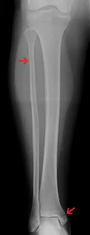 | |
| A Maisonneuve fracture with arrows marking the location of the two fractures | |
| Specialty | Orthopedics |
| Symptoms | Tenderness of medial ankle and outside aspect of the upper part of the lower leg[1] |
| Complications | Osteoarthritis[2] |
| Causes | Forceful, external rotation of the foot[3] |
| Diagnostic method | X-ray[4] |
| Differential diagnosis | Isolated posterior malleolus facture[4] |
| Treatment | Surgery[3] |
| Frequency | Relatively uncommon[1] |
A Maisonneuve fracture is an injury at the ankle with an associated break of the lower leg near the knee.[5] The ankle injury involves a tear of the ligament connecting the two bones of the lower leg, the tibiofibular syndesmosis.[5] There may also be a break of the medial malleolus or rupture of the deltoid ligament of the ankle.[3] Symptoms typically include tenderness over the medial ankle and outside aspect of the upper part of the lower leg.[1]
It typically results from excessive external rotation of the ankle.[3] X-rays typically show widening of the ankle joint, though this may be subtle.[3][1] Stress views may be useful in unclear cases.[4] People may not mention pain around the knee due to the greater degree of pain in the ankle.[6] It is classified as a type C3 ankle fracture according to the Danis-Weber classification system.[7]
Treatment is generally by surgery, including placing screws to hold together the two bones of the lower leg.[3] These screws may be removed after two to three months.[1] With treatment most people have good outcomes.[2] Maisonneuve fractures are relatively uncommon.[1] Males are more commonly affected than females.[4] They are named after the surgeon Jules Germain François Maisonneuve, who first describe it in the 1800s.[3][8]
Signs and symptoms
Common symptoms are pain, swelling, tenderness, and bruising around the ankle joint and inferior (or distal) tibiofibular joint. More specifically, as a pronation-external rotation injury, pain during external rotation of the ankle joint is expected. Additionally, there is a reduced range of motion of the foot and an inability to weight-bear due to ankle pain.[9][10] Pain may also be felt around the medial and lateral aspects of the ankle, and more rarely around the superior (or proximal) tibiofibular joint.[11] Damage to the deltoid ligament or interosseous membrane can cause bleeding around the surrounding tissues, resulting in a localised oedema.[12]
Complications
As the syndesmotic ligaments are responsible for stabilising the ankle mortise and tibiotalar joint, disruption to this syndesmosis can cause a reduction of the space between the distal tibia, fibula, and talus. A long-term effect of this is painful ankle osteoarthritis due to the direct contact between the tibia and talus.[13][14]
If left untreated, instability of the tibiotalar joint and deltoid ligament can cause a valgus deformity of the ankle. This leaves the ankle joint in a state of chronic pronation, characterised by a protrusion of the medial malleolus into the subcutaneous tissue.[14]
Cause
Forceful, external rotation of the ankle joint is the main cause of a Maisonneuve fracture.[9][10] Engaging in high-intensity sports or falling over can increase the risk of tearing the deltoid ligament or cause an avulsion fracture of the medial malleolus from external rotation of the foot.[9][8] In some cases, motor vehicle accidents can also result in a Maisonneuve fracture.[12]
Pathophysiology
The Maisonneuve fracture generally follows a specific pattern of injury. The following are described:[15][9][16]
- Forceful, external rotation of the ankle joint results in the tearing of the deep deltoid ligament and/or an avulsion fracture of the medial malleolus.
- The ankle mortise is subjected to excessive torque, rupturing the syndesmotic ligaments and anteromedial ankle joint capsule.
- Rotative energy is transferred upwards along the interosseous membrane, damaging it in the process.
- The force results in a spiral, sometimes an oblique, fracture at the neck of the proximal fibula.
In cases where the anterior aspect of the tibiofibular syndesmosis can resist mechanical stress, only an oblique fracture of the lateral malleolus is produced. Diastasis of the lateral malleolus may also occur, in which it is posterolaterally displaced from the tibia.[11]
Although most Maisonneuve cases report a pronation-external rotation mechanism of injury, clinical studies have recorded instances of supination-external rotation being the mechanism of injury.[8] Slight or high degrees of plantarflexion prior to supination-external rotation of the foot have been identified in patients with proximal fibular fractures.[12]
The Maisonneuve fracture is similar to the Galeazzi fracture in the sense that there is an important ligamentous disruption in association with the fracture.[17]
Diagnosis
Diagnosing requires a combination of medical history, physical examination, and radiographic imaging.[14] People generally do not report pain near the proximal fibula, so examination such as palpation along the fibula is effective for differentiating a Maisonneuve fracture from an isolated syndesmotic injury.[9] Feeling pain near the proximal fibula during palpation is a possible indication of a Maisonneuve fracture.[16] Ankle instability is often associated with a damaged proximal fibula in a Maisonneuve fracture, so people are typically asked about the mechanism of injury. Mortise stability is examined to rule out the possibility of an isolated fibular fracture.[8]
Ankle Xrays are used to detect widening of the tibiofibular syndesmosis or medial clear space. The medial clear space is the area between the talus of the ankle and the medial malleolus. Damage to the deltoid ligament and syndesmotic ligaments result in mortise instability, causing the talus to laterally shift and widen the medial clear space.[9][16] A study found that the medial clear space size of a normal ankle and an injured ankle measured at 4 millimetres and 5.4 millimetres in length respectively.[14] To confirm diagnosis, full-leg radiographs are used to inspect for fractures of the proximal fibula and widening of the interosseous clear space (or tibiofibular clear space). The interosseous clear space is the area between the medial side of the fibula and lateral side of the tibia. A peer-reviewed study, published in Injury in 2004, found that an interosseous clear space greater than 10 millimetres indicates diastasis of the syndesmotic ligaments.[9]
If necessary, CT scan or magnetic resonance imaging may also be used to clarify diagnosis. MRI scans can check for interosseous membrane or tibial tubercle damage if high instability of the ankle is diagnosed.[12][14] Arthroscopy may be used to diagnose a syndesmotic lesion but is often not recommended due to operative difficulty.[18] Stress radiographs of the ankle are used to assess the integrity of the deltoid ligament and tibiofibular syndesmosis.[11][18] The size of the medial clear space can also be measured using stress radiography.[14]
Classification
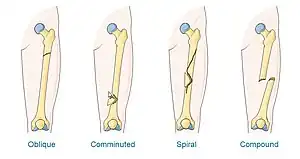
A Maisonneuve fracture may be a simple fracture or comminuted fracture:[12][19]
- A simple fracture, in the case of a Maisonneuve fracture, refers to the fibula being broken in one place without any damage being done to the surrounding tissues.
- A comminuted fracture is when the bone is broken in more than two places.
X-ray, CT, or MRI can be used to diagnose the extent of damage and determine whether it is a simple or comminution fracture.[12] During diagnosis, a supination-external rotation pattern of injury may also be concluded if there is an isolated fracture of the posterior tubercle of the tibia.[11]
Treatment
- Reduce the proximal fibula and medial malleolus to achieve stabilisation
- Repair the distal tibiofibular syndesmosis and deltoid ligament
- Restore ankle mortise stability
Treatment can be achieved by either non-operative (or conservative) or operative means. The main operative treatments for a Maisonneuve fracture are open-reduction surgery and closed-reduction surgery, both of which usually preceding internal fixation of the injury. These procedures are known as Open Reduction Internal Fixation (ORIF) and Closed Reduction Internal Fixation (CRIF).[8][18]
Surgery
Syndesmotic screws are the main, internal fixators used in a Maisonneuve fracture. Two main types of syndesmotic screws are used: trans-syndesmotic screws (positioned at the level of the syndesmosis) and supra-syndesmotic screws (positioned above the syndesmosis).[20]
Syndesmotic screws are recommended to be fixed at least 1 centimetre proximal to the tibiofibular syndesmosis or 4 to 6 centimetres proximal to the tibiotalar joint line.[9][21] Cadaveric analyses suggest that screw fixation at 2 centimetres proximal to the tibiotalar joint line is also adequate.[22] Biodegradable implants such as bioabsorbable screws, which do not require postoperative removal, may be used as an alternative to metallic hardware. However, biodegradable implants still limit rotation of the ankle and dorsiflexion of the foot.[9][8][18]
Open reduction
Open-reduction surgery is typically not performed at the level of the proximal fibula, as dissection near the proximal end may risk severing the common peroneal nerve. Instead, reducing the proximal fibula at the level of the distal tibiofibular syndesmosis is recommended.[8][16] A hook test is performed, using a curved hook, to assess the stability of the fibula. If instability is detected, further distraction of the fibula can be done to repair the full bone. The fibula can then be guided into the fibular notch located on the tibia, effectively restoring its length. Internal rotation of the foot may then be used to correct anatomical alignment.[16][18]
Following open-reduction, internal fixation is usually performed to stabilise the ankle mortise. To account for the distal fibula being slightly posterior to the distal tibia, drill holes are angled at 30° from the anteromedial aspect of the tibia to the posterolateral aspect of the fibula.[9][16][21] Trans-syndesmotic screws can be inserted in this way to ensure tibia fixation. Additional supra-syndesmotic screws may be temporarily inserted, for approximately 3 to 6 months, if instability is still present after fibular reduction. To reduce the fibula and restore the ankle mortise to its proper anatomical configuration, partial dorsiflexion of the foot is maintained prior to intraoperative screw fixation. This is because, in a neutral or maximally dorsiflexed position of the foot, the trochlear surface of the talus may reduce maximal postoperative dorsiflexion due to rigidity after screw fixation.[11][16]
Assessing the severity of syndesmotic lesions can be performed with fluoroscopic screening.[18] Guidance under fluoroscopy can also assist with syndesmotic screw fixation.[9] Restoration of the anteromedial joint capsule of the ankle can be achieved with suturing techniques.[10][11]
Closed reduction
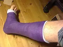
Closed-reduction surgery requires no dissection or incisions being made into the leg to operate. It is most commonly applied in cases where the Maisonneuve fracture has only damaged the anterior portion of the syndesmotic ligaments.[8] That is, the posterior hinge of the ankle is still stable, and the foot can be internally rotated using traction to restore fibular bone length.[9] Long-leg casting or short-leg casting is applied postoperatively to maintain this alignment.[16]
It is generally recommended that medial malleolar fractures do not require surgical intervention if closed reduction is sufficient for the restoration of bone length. Otherwise, large medial malleolar fractures can be fixed using trans-syndesmotic screws, figure-of-8 wires, or Kirschner wires.[8] For smaller medial malleolar fractures, repair with a wire-tension band is sufficient.[11]
No surgery
In cases where only the posterior ligaments of the tibiofibular syndesmosis are partially damaged, non-operative treatment such as long-leg casting for at least 6 weeks is recommended.[9][23] Immobilisation such as casting are often paired with non-weight bearing precautions.[14][16] Gradually, physiotherapy rehabilitation programs allow people to weight-bear after at least 8 weeks of postoperative casting.[16] These non-operative treatments may also be used in cases where the medial malleolus remains intact.[8]
Complications
Delaying diagnosis and treatment can result in intraoperative complications. In one case the insertion of a super-syndesmotic screw caused the lateral malleolus to be further shifted laterally; subsequent removal of the screw was necessary.[10]
Complications that may postoperatively occur include:
- the development of osteoarthritis around the ankle joint.[14]
- the development of peroneal nerve palsy following damage to the common peroneal nerve.[16]
- extra-osseous ("outside of the bone") calcification of vessels or tissues surrounding the tibiofibular joint.[10]
Hardware
Incorrectly positioned screws can potentially make contact with articular surfaces, which can cause calcification around the affected area. Screw breakage can also cause pain in these areas.[9]
Postoperative hardware removal can cause problems such as infection, joint rigidity, or diastasis if fixation was not sufficiently long enough.[18] In areas where residual stiffness has persisted, patients may report feeling pain or a mild aching sensation.[11] Generally, it is recommended that hardware removal should be done anywhere from 6 weeks to 12 weeks after internal fixation to allow the tibiofibular syndesmosis to properly heal.[8][16] Syndesmotic screws should be removed prior to rehabilitative training; bearing weight without prior hardware removal may result in ankle stiffness due to reduced dorsiflexion of the foot and potential screw breakage.[9]
Follow-ups
Postoperative follow-ups are done to ensure that treatment has produced satisfactory results, such as checking if malreduction of any of the associated structures occured.[18][24] Follow-ups may be performed from 6 months to 2 years after the surgery and are applicable for both non-operative and operative treatments.[9][11]
Epidemiology
Exact rates are unknown, but it is believed that the Maisonneuve fracture accounts for 5% of all ankle injuries treated in surgery.[11][23] The Maisonneuve fracture has been reported in people as young as 17–19 years old, and up to 42–70 years old.[10][11][14] The injury is mostly seen in males, making up about 78% of cases.[12]
Sporting injuries are the most common risk factor, representing about 50% of cases.[9]
References
- 1 2 3 4 5 6 Thordarson, DB (September 1996). "Detecting and treating common foot and ankle fractures. Part 1: the ankle and hindfoot". The Physician and sportsmedicine. 24 (9): 29–38. doi:10.3810/psm.1996.09.1337. PMID 20087018.
- 1 2 Porter, DA; Jaggers, RR; Barnes, AF; Rund, AM (2014). "Optimal management of ankle syndesmosis injuries". Open access journal of sports medicine. 5: 173–82. doi:10.2147/OAJSM.S41564. PMID 25177153.
- 1 2 3 4 5 6 7 Weerakkody, Yuranga. "Maisonneuve fracture | Radiology Reference Article | Radiopaedia.org". Radiopaedia. Archived from the original on 2 September 2022. Retrieved 3 April 2023.
- 1 2 3 4 Bennett, D. Lee; El-Khoury, Georges Y. (23 May 2013). Pearls and Pitfalls in Musculoskeletal Imaging: Variants and Other Difficult Diagnoses. 135: Cambridge University Press. ISBN 978-1-107-06700-4. Archived from the original on 3 April 2023. Retrieved 3 April 2023.
{{cite book}}: CS1 maint: location (link) - 1 2 Natrajan, M. V. (2018). "20. Injuries of the ankle and foot". Natarajan’s Textbook of Orthopaedics & Traumatology (8th ed.). New Delhi: Wolters Kluwer. p. 334-335. ISBN 978-93-86691-49-1. Archived from the original on 2022-09-15. Retrieved 2022-09-14.
- ↑ Essentials of skeletal radiology. Lippincott Williams & Wilkins. 2005. p. 538. Archived from the original on 2023-04-03. Retrieved 2023-04-03.
- ↑ Gaillard, Frank. "Weber classification of ankle fractures | Radiology Reference Article | Radiopaedia.org". Radiopaedia. Archived from the original on 13 January 2023. Retrieved 3 April 2023.
- 1 2 3 4 5 6 7 8 9 10 11 Stufkens, SA; van den Bekerom, MP; Doornberg, JN; van Dijk, CN; Kloen, P (January 2011). "Evidence-based treatment of maisonneuve fractures". The Journal of foot and ankle surgery : official publication of the American College of Foot and Ankle Surgeons. 50 (1): 62–7. doi:10.1053/j.jfas.2010.08.017. PMID 21172642. Archived from the original on 2021-09-11. Retrieved 2022-08-23.
- 1 2 3 4 5 6 7 8 9 10 11 12 13 14 15 16 17 Sproule, J. A., Khalid, M., O’Sullivan, M., & McCabe, J. P. (2004). Outcome after surgery for Maisonneuve fracture of the fibula. Injury. 35(8): 791-798. Archived 2021-09-11 at the Wayback Machine
- 1 2 3 4 5 6 Babis, G. C., Papagelopoulos, P. J., Tsarouchas, J., Zoubos, A. B., Korres, D. S., & Nikiforidis, P. (2000). Operative treatment for maisonneuve fracture of the proximal fibula. Orthopedics. 23(7): 687-690. Archived 2021-09-14 at the Wayback Machine
- 1 2 3 4 5 6 7 8 9 10 11 Pankovich, A. M. (1976). Maisonneuve fracture of the fibula. J Bone Joint Surg Am. 58(3): 337-342. Archived 2020-11-06 at the Wayback Machine
- 1 2 3 4 5 6 7 He, J., Ma, X., Xin, J., Cao, H., Li, N., Sun, Z., Wang, G., Fu, X., Zhao, B., & Hu, F. (2020). Pathoanatomy and Injury Mechanism of Typical Maisonneuve Fracture. Orthopaedic Surgery. DOI: 10.1111/os.12733 Archived 2021-09-14 at the Wayback Machine
- ↑ Ramsey, P. L. & Hamilton, W. (1976). Changes in tibiotalar area of contact caused by lateral talar shift. J Bone Joint Surg Am. 58(3): 356-357. Archived 2021-02-26 at the Wayback Machine
- 1 2 3 4 5 6 7 8 9 Levy, B. A., Vogt, K. J., Herrera, D. A., & Cole, P. A. (2006). Maisonneuve fracture equivalent with proximal tibiofibular dislocation. A case report and literature review. J Bone Joint Surg Am. 88(5): 1111-1116. Archived 2021-09-11 at the Wayback Machine
- ↑ Lauge-Hansen, N. (1950). Fractures of the ankle. II. Combined experimental-surgical and experimental-roentgenologic investigations. Arch Surg. 60(5): 957- 985. Archived 2020-06-20 at the Wayback Machine
- 1 2 3 4 5 6 7 8 9 10 11 12 Duchesneau, S. & Fallat, L. M. (1995). The Maisonneuve Fracture. J Foot Ankle Surg. 34(5): 422-428. Archived 2020-11-08 at the Wayback Machine
- ↑ Atesok, K. I., Jupiter, J. B., & Weiss, A. P. (2011). Galeazzi fracture. J Am Acad Orthop Surg. 19(10): 623-633. Archived 2020-11-08 at the Wayback Machine
- 1 2 3 4 5 6 7 8 Schnetzke, M., Vetter, S. Y., Beisemann, N., Swartman, B., Grützner, P. A., & Franke, J. (2016). Management of syndesmotic injuries: What is the evidence?. World J Orthop. 7(11): 718-725. Archived 2020-11-06 at the Wayback Machine
- ↑ Giorgi, Anna (8 July 2017). "Fracture". Healthline. Archived from the original on 7 September 2021. Retrieved 23 August 2022.
- ↑ Kukreti, S., Faraj, A., & Miles, J. N. V. (2005). Does position of syndesmotic screw affect functional and radiological outcome in ankle fractures?. Injury. 36(9): 1121-1124. Archived 2022-09-15 at the Wayback Machine
- 1 2 van den Bekerom, M. P. J. & Raven, E. E. J. (2007). Current concepts review: operative techniques for stabilizing the distal tibiofibular syndesmosis. Foot Ankle Int. 28(12): 1302-1308. Archived 2021-09-11 at the Wayback Machine
- ↑ McBryde A., Chiasson, B., Wilhelm, A., Donovan, F., Ray, T., & Bacilla, P. (1997). Syndesmotic screw placement: a biomechanical analysis. Foot Ankle Int. 18(5): 262-266. Archived 2021-09-11 at the Wayback Machine
- 1 2 Lock, T.R., Schaffer, J. J., & Manoli, A. (1987). Maisonneuve fracture: case of missed diagnosis. Ann Emerg Med. 16(7): 805-807. Archived 2021-09-11 at the Wayback Machine
- ↑ Pietrangelo, Ann (7 July 2018). "Postoperative Care". Healthline. Archived from the original on 1 June 2022. Retrieved 23 August 2022.
External links
| Classification | |
|---|---|
| External resources |