Osteoma
| Osteoma | |
|---|---|
| Other names: Benign osteogenic tumors, unspecified and osteoma NOS.[1] Not recommended: ivory exostosis, parosteal osteoma, torus palatines/mandibularis[1] | |
 | |
| CT scan showing an osteoma growing on inside of skull bone | |
| Specialty | Orthopedics |
| Symptoms | None, pain, headache[1] |
| Complications | Pressure symptoms[1] |
| Usual onset | Slow[1] |
| Causes | Unknown[1] |
| Diagnostic method | Medical imaging: X-ray, CT scan, MRI[1] |
| Treatment | None, surgery[1] |
| Frequency | Males=Females[1] |
Osteoma (plural: "osteomata" or "osteomas"), is a non-cancerous bone tumor, a type osteogenic tumor, where a new piece of bone typically grows on another piece of bone, usually on the skull and near the sinuses.[1][2] Often there are no symptoms as the tumor grows slowly, but there may be pain, headache, blocked paranasal sinuses or local swelling.[1] It may present with sinusitis.[3]
The cause is unknown.[1] Osteomas are also found in Gardner's syndrome.[1]
Medical imaging such as X-ray, CT scan and MRI show dense, clearly defined, round white tumors attached to bone.[1] They can be left alone if not troubling, and surgically cut out if pressure symptoms.[1] The surgery may be possible through the nose, without making a large cut.[3]
It affects males and females equally.[1] How commonly it occurs is not known but some report up to 6.4% may be affected.[1] Findings of osteomata date back to ancient Egypt (664-332 BCE).[1]
Signs and symptoms

There may be no symptoms or blocked paranasal sinuses or local swelling.[1] An affected person might therefore present with an infection of the sinuses, headaches or facial pain.[3]
Often, craniofacial osteoma presents itself through eye signs and symptoms (such as protruding eyes).[4]
Cause
The cause of osteomas is uncertain, but commonly accepted theories propose congenital, traumatic, or infectious causes.[3] Osteomas are also found in Gardner's syndrome.[1]
Diagnosis
Medical imaging such as X-ray, CT scan and MRI show dense, clearly defined, round white tumors attached to bone.[1] They may be diagnosed when having medical imaging for another reason.[3] Osteomas of the paranasal sinuses and skull base can be diagnosed using CT-scan without intravenous contrast, allowing its size and relation to nearby important structures to be assessed.[3] A biopsy is not usually required.[3]
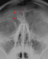 X-ray skull: Osteoma of the frontal sinus
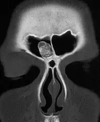 CT-scan skull: Osteoma of the frontal sinus
.JPEG.webp) X-ray skull: Osteoma of the frontal sinus
X-ray skull: Osteoma of the frontal sinus
Treatment
They can be left alone if not troubling, and surgically cut out if pressure symptoms.[1]
Epidemiology
It most frequently affects the skull and affect males and females equally.[1] The true prevalence is not known but some reports quote up to 6.4%.[1]
Osteoma represents the most common non-cancerous tumor of the nose and paranasal sinuses.[3]
History
Findings of osteomata date back to ancient Egypt (664-332 BCE).[1]
Variants
- Osteoma cutis
- Fibro-osteoma
- Chondro-osteoma
See also
References
- 1 2 3 4 5 6 7 8 9 10 11 12 13 14 15 16 17 18 19 20 21 22 23 24 25 WHO Classification of Tumours Editorial Board, ed. (2020). "Bone tumors". Soft Tissue and Bone Tumours: WHO Classification of Tumours. Vol. 3 (5th ed.). Lyon (France): International Agency for Research on Cancer. p. 391-393. ISBN 978-92-832-4503-2. Archived from the original on 2021-06-13. Retrieved 2021-07-06.
- ↑ "ICD-11 - ICD-11 for Mortality and Morbidity Statistics". icd.who.int. Archived from the original on 1 August 2018. Retrieved 16 July 2021.
- 1 2 3 4 5 6 7 8 "Osteoma". Stanford Medicine: Skull Base Surgery (in Gagana Samoa). Archived from the original on 18 January 2021. Retrieved 15 July 2021.
- ↑ Michael S. Schwartz, MD; Dennis M. Crockett, MD. "Management of a Large Frontoethmoid Osteoma with Sinus Cranialization and Cranial Bone Graft Reconstruction". International Journal of Pediatric Otorhinolaryngology. Archived from the original on 2012-02-16. Retrieved 2020-03-24.
External links
| Classification |
|---|