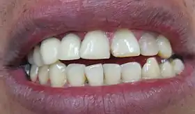
Tooth resorption
| Root resorption | |
|---|---|
| Specialty | Dentistry |
Tooth resorption, or root resorption, is the progressive loss of dentine and cementum by the action of osteoclasts.[1] This is a normal physiological process in the exfoliation of the primary dentition, caused by osteoclast differentiation due to pressure exerted by the erupting permanent tooth. However, in the secondary dentition the process is pathological.
Cause
Pulp necrosis, trauma, periodontal treatment, orthodontics and tooth whitening are the most common stimulants of inflammatory resorption.[2] Some other less common causes include pressure from ectopic teeth, cysts, and tumours. These cause damage to the periodontal ligament (PDL), cementum, or pre-dentine. These tissues provide protection from resorption when intact and healthy.
Regardless of the reason, tooth resorption is due to an inflammatory process in response to an insult on one of the above.[3][4]
Mechanism
Osteoclasts are the cells responsible for the resorption of the root surface.[5] Osteoclasts can break down bone, cartilage and dentine.
Receptive activator of nuclear factor kappa-B ligand (RANKL), also called osteoclast differentiation factor (ODF) and osteoprotegerin ligand (OPGL), is a regulator of osteoclast function.[6][7] In physiological bone turn over, osteoblasts and stromal cells release RANKL, this acts on macrophages and monocytes which fuse and become osteoclasts.[8] Osteoprotegerin (OPG) is also secreted by osteoclasts and stromal cells; this inhibits RANKL and therefore osteoclast activity.
The pathophysiology of stimulation of osteoclasts in the process of inflammatory resorption is unknown.
One thought is that the presence of bacteria plays a role. Bacterial presence leads to pulpal or peri-periapical inflammation. These bacteria are not mediators of osteoclast activity but do cause leukocyte chemotaxis. Leukocytes differentiate into osteoclasts in the presence of lipopolysaccharide antigens found in Porphyromonas, Prevotella and Treponema species (these are all bacterial species associated with pulpal or periapical inflammation).[9]
Osteoclasts are active during bone regulation, there is constant equilibrium of bone resorption and deposition. Damage to the periodontal ligament can lead to RANKL release activating osteoclasts.[10] Osteoclasts in close proximity to the root surface will resorb the root surface cementum and underlying root dentin. This can vary in severity from evidence of microscopic pits in the root surface to complete devastation of the root surface.
When there is insult leading to inflammation (trauma, bacteria, tooth whitening, orthodontic movement, periodontal treatment) in the root canal/s or beside the external surface of the root, cytokines are produced, the RANKL system is activated and osteoclasts are activated and resorb the root surface.
If the insult is transient, resorption will stop and healing will occur, this is known as transient inflammatory resorption.[11] If the insult is persistent, then resorption continues, and if the tooth tissue is irretrievably damaged, complete resorption may occur.[12]
Classifications
Internal resorption
Internal resorption defines the loss of tooth structure from within the root canal/s of a tooth.
It may present initially as a pink-hued area on the crown of the tooth; the hyperplastic, vascular pulp tissue filling in the resorbed areas. This condition is referred to as a pink tooth of Mummery, after the 19th century anatomist John Howard Mummery. It may also present as an incidental, radiographic finding. Radiographically a radiolucent area within the root canal may be visible and/or the canal may appear sclerosed.
Chronic pulpal inflammation is thought to be a cause of internal resorption. The pulp must be vital below the area of resorption to provide osteoclasts with nutrients. If the pulp becomes totally necrosed the resorption will cease unless lateral canals are present to supply osteoclasts with nutrients.
If the condition is discovered before perforation of the crown or root has occurred, endodontic therapy (root canal therapy) may be carried out with the expectation of a fairly high success rate. Removing the stimulus (inflamed pulp) results in cessation of the resorptive process.
External resorption

External resorption is the loss of tooth structure from the external surface of the tooth. It can be further divided in the following classifications:[4]
Inflammatory
External inflammatory resorption occurs following prolonged insult leading to continuing pathological resorption. It is commonly caused by damage to the periodontal ligament (PDL), drying of root surface following avulsion, exposure of dentine tubules, and pressure. This process can occur rapidly.
Surface
Also known as transient inflammatory resorption. It is a self-limiting process and is a often and incidental radiographic finding. Transient inflammatory resorption undergoes healing and should be monitored only.
It is caused by localised and limited injury to root surface or surrounding tissues.[11][13] There is 2–3 weeks of osteoclast activity before healing then occurs. If cementum alone is involved in the resorptive process then complete healing will occur, but if dentine is involved there will be re-contouring in the area of lost dentine.[11]
Cervical
External cervical resorption is a localised resorptive lesion in the cervical area of the tooth, below the epithelial attachment. This rarely involves the pulp. Prolonged insult leads to vertical and horizontal growth of the lesion. It is commonly caused by trauma, periodontal treatment, or tooth whitening.
Multiple idiopathic cervical resorption is when a minimum of 3 teeth are affected by cervical resorption for no evident cause.
Replacement

External replacement resorption occurs following ankylosis of the root of the alveolar bone. The tooth tissue is resorbed and replaced with bone. This process is poorly understood.
It is thought that following the union of bone and tooth and the obliteration of the PDL, the protective regulators released by the PDL to protect the root from resorption are no longer present. This results in the tooth tissue being resorbed by osteoclasts and replaced with bone like it was part of the continuous homeostatic process of bone turnover.
There is currently no single best intervention or treatment for the management of external root resorption. Treatments are usually case-dependent and therefore more research is needed in this area. [14]
See also
- Cementoblastoma
- Tooth ankylosis
- Feline odontoclastic resorptive lesion
References
- ↑ Patel S, Ford TP (May 2007). "Is the resorption external or internal?". Dental Update. 34 (4): 218–20, 222, 224–6, 229. doi:10.12968/denu.2007.34.4.218. PMID 17580820.
- ↑ Fuss Z, Tsesis I, Lin S (August 2003). "Root resorption--diagnosis, classification and treatment choices based on stimulation factors". Dental Traumatology. 19 (4): 175–82. doi:10.1034/j.1600-9657.2003.00192.x. PMID 12848710.
- ↑ Andreasen JO (April 1985). "External root resorption: its implication in dental traumatology, paedodontics, periodontics, orthodontics and endodontics". International Endodontic Journal. 18 (2): 109–18. doi:10.1111/j.1365-2591.1985.tb00427.x. PMID 2860072.
- 1 2 Darcey J, Qualtrough A (May 2013). "Resorption: part 1. Pathology, classification and aetiology". British Dental Journal. 214 (9): 439–51. doi:10.1038/sj.bdj.2013.431. PMID 23660900.
- ↑ Boyle WJ, Simonet WS, Lacey DL (May 2003). "Osteoclast differentiation and activation". Nature. 423 (6937): 337–42. Bibcode:2003Natur.423..337B. doi:10.1038/nature01658. PMID 12748652. S2CID 4428121.
- ↑ Yasuda H, Shima N, Nakagawa N, Yamaguchi K, Kinosaki M, Mochizuki S, et al. (March 1998). "Osteoclast differentiation factor is a ligand for osteoprotegerin/osteoclastogenesis-inhibitory factor and is identical to TRANCE/RANKL". Proceedings of the National Academy of Sciences of the United States of America. 95 (7): 3597–602. Bibcode:1998PNAS...95.3597Y. doi:10.1073/pnas.95.7.3597. PMC 19881. PMID 9520411.
- ↑ Mak TW, Saunders ME (2006). "Cytokines and Cytokine Receptors". The Immune Response. Elsevier: 463–516. doi:10.1016/b978-012088451-3.50019-3. ISBN 978-0-12-088451-3.
- ↑ Nakamura I, Takahashi N, Jimi E, Udagawa N, Suda T (April 2012). "Regulation of osteoclast function". Modern Rheumatology. 22 (2): 167–77. doi:10.3109/s10165-011-0530-8. PMID 21953286. S2CID 218989802.
- ↑ Choi BK, Moon SY, Cha JH, Kim KW, Yoo YJ (May 2005). "Prostaglandin E(2) is a main mediator in receptor activator of nuclear factor-kappaB ligand-dependent osteoclastogenesis induced by Porphyromonas gingivalis, Treponema denticola, and Treponema socranskii". Journal of Periodontology. 76 (5): 813–20. doi:10.1902/jop.2005.76.5.813. PMID 15898943.
- ↑ Trope M (2002). "Root resorption due to dental trauma". Endodontic Topics. 1: 79–100. doi:10.1034/j.1601-1546.2002.10106.x.
- 1 2 3 Andreasen JO (1981). "Relationship between cell damage in the periodontal ligament after replantation and subsequent development of root resorption. A time-related study in monkeys". Acta Odontologica Scandinavica. 39 (1): 15–25. doi:10.3109/00016358109162254. PMID 6943905.
- ↑ Lossdörfer S, Götz W, Jäger A (July 2002). "Immunohistochemical localization of receptor activator of nuclear factor kappaB (RANK) and its ligand (RANKL) in human deciduous teeth". Calcified Tissue International. 71 (1): 45–52. doi:10.1007/s00223-001-2086-7. PMID 12043011. S2CID 20902759.
- ↑ Majorana A, Bardellini E, Conti G, Keller E, Pasini S (October 2003). "Root resorption in dental trauma: 45 cases followed for 5 years". Dental Traumatology. 19 (5): 262–5. doi:10.1034/j.1600-9657.2003.00205.x. PMID 14708650.
- ↑ Ahangari, Zohreh; Nasser, Mona; Mahdian, Mina; Fedorowicz, Zbys; Marchesan, Melissa A. (2015-11-24). "Interventions for the management of external root resorption". The Cochrane Database of Systematic Reviews (11): CD008003. doi:10.1002/14651858.CD008003.pub3. ISSN 1469-493X. PMC 7185846. PMID 26599212.
