This article was medically reviewed by Luba Lee, FNP-BC, MS. Luba Lee, FNP-BC is a Board-Certified Family Nurse Practitioner (FNP) and educator in Tennessee with over a decade of clinical experience. Luba has certifications in Pediatric Advanced Life Support (PALS), Emergency Medicine, Advanced Cardiac Life Support (ACLS), Team Building, and Critical Care Nursing. She received her Master of Science in Nursing (MSN) from the University of Tennessee in 2006.
This article has been viewed 23,823 times.
The optic nerve connects the back of the eyeball to the cerebrum, and transmits visual perceptions to your brain. Testing the optic nerve is a routine part of regular checkups at your primary physician’s office or at your optometrist’s office. During a full optic-nerve exam, your optometrist will examine your visual abilities and reflexes in order to confirm that your nerve is functioning properly and your eyes pick up visual information correctly. The doctor will also shine a light into your eyes in order to visually inspect the shape and alignment of your pupils.
Steps
Testing Visual Acuity
-
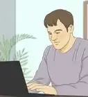
1Position yourself about 6 metres (20 ft) from the Snellen chart. The Snellen chart is the large chart found in all doctor’s offices which features the letters of the alphabet, arranged in 8 rows at random, in decreasing size.[1]
- In most optic-nerve exams, the optometrist or an assistant will direct you where to stand or sit.
-
2Cover one of your eyes with the palm of your hand. The Snellen chart is meant to be read with only one eye at a time, in order to test the acuity of each eye individually. In some cases, the doctor may provide a plastic spoon-like utensil that you can use to cover your eye. Otherwise, cover the eye completely with the palm of your hand.[2]
- If you routinely wear glasses or contact lenses, keep them on for the exam unless the doctor directs you to do otherwise.
- Visual acuity is a numerical value derived by as your distance from the chart over the number of the lowest line that you correctly read. For example, 20/20 (or 6/6, using meters) is perfect vision.
Advertisement -
3Read the lowest line that you’re able to on the Snellen chart. Lower lines on the chart feature smaller letters, meaning that you’ll most likely be unable to read the lowest 2 or 3 lines. Pick a line on the bottom half of the chart, and read the letters as best you can.[3]
- Following this reading, the doctor may ask you to attempt to read a higher or lower line on the chart.
- A line is considered to have been successfully read if you misread 2 or fewer letters.
-
4Repeat the steps with your other eye. Once you’ve read the lowest possible line with one eye, remove your hand and use it to cover your other eye. Then begin the process over again by attempting to read a low line on the Snellen chart with your second eye covered.[4]
- Once you’ve finished the acuity exam with your second eye, you can ask the doctor what your acuity score was.


Testing Visual Fields
-
1Remain still and look straight ahead at the optometrist. For the visual field test, it’s important that you keep your eyes focused directly ahead. The doctor will stand about 3 feet (0.91 m) in front of you. They will hold out their hand about 1 foot (0.30 m) from one side of your face, and wiggle one of their fingers at the same level as your eyes. They’ll ask you to confirm that you saw the finger wiggle.
- Testing your visual field involves making sure that your optic nerve correctly picks up and transmits visual data from your peripheral vision. It also determines if there are any lesions in your visual pathway.
-
2Keep your eyes ahead while the doctor repeats the procedure. For a full visual field exam, the optometrist will test your optic nerve’s ability to gather visual data in each of the four quadrants of your peripheral vision: upper-right, upper-left, lower-right, and lower-left. The doctor will repeat the finger-wiggling exercise three times, and ask you to confirm that you saw the movement of their finger.
- If at any point you cannot see motion in your peripheral vision, let the doctor know. This may indicate a problem with your optic nerve.
-
3Ask the doctor about a visual inattention test. This is a further means of testing your optic nerve’s ability to pick up information in your peripheral vision. In a visual inattention test, the optometrist will hold out their hands as in a typical visual field exam, but they’ll wiggle more than one finger simultaneously.
- The doctor will ask you to identify how many fingers they wiggled and which specific fingers they were.


Testing Visual Reflexes
-
1Allow the optometrist to shine a penlight into your eye. The doctor will place one of their hands vertically between your eyes, and then shine the light directly into one of your pupils to check your eyes' alignment. The hand placement ensures that the light doesn’t cause the pupil of your second eye to dilate while the doctor is testing your first eye.
- You may experience minor discomfort from the bright pen light during this procedure. However, the light won’t harm your eye.
-
2Look straight ahead into the light. Don’t avert your eyes from the light, and let your pupils dilate naturally. The optometrist needs to observe your pupil dilation. If your pupils do not dilate, or if they constrict slowly, there may be a problem with one or both of your optic nerves.[5]
-
3Repeat the process with your other eye. The doctor will keep their hand held vertically between your eyes, and shine the penlight into your other pupil. As with your first eye, they'll look to make sure that your pupil dilates fully.
- Once the visual reflex exam is complete, you can ask the doctor if your eyes dilated as expected.


Assessing Ocular Movement
-
1Ask about a cover test. Cover tests can be used by your optometrist to determine if your eyes are deviated, or in strabismus evaluation. In this exam, your optometrist will ask you to look straight ahead at a target. They will then cover one eye with a card. If your doctor notices movement in the uncovered eye, you may have some deviation. The test is then repeated on the other eye.[6]
- Your doctor will also check your covered eye as they uncover it. If the covered eye does not move, no strabismus is present.
-
2Let your doctor evaluate 6 positions of your gaze. Weak or paralyzed extraocular eye muscles can cause a deviated eye. Your optometrist will be able to check for this by directing you to look at 6 cardinal points of gaze. Simply follow their instructions and allow them to observe your eye during this process.[7]


References
- ↑ https://www.ucsfhealth.org/medical-tests/visual-acuity-test
- ↑ https://www.ucsfhealth.org/medical-tests/visual-acuity-test
- ↑ https://geekymedics.com/cranial-nerve-exam/
- ↑ https://geekymedics.com/cranial-nerve-exam/
- ↑ https://geekymedics.com/cranial-nerve-exam/
- ↑ https://webeye.ophth.uiowa.edu/eyeforum/video/basic-cover-test.htm
- ↑ https://www.dartmouth.edu/~dons/part_1/chapter_4.html
- ↑ https://geekymedics.com/cranial-nerve-exam/
- ↑ https://www.glaucoma.org/treatment/imaging-of-the-optic-nerve-what-is-it-and-why-is-it-needed.php


Medical Disclaimer
The content of this article is not intended to be a substitute for professional medical advice, examination, diagnosis, or treatment. You should always contact your doctor or other qualified healthcare professional before starting, changing, or stopping any kind of health treatment.
Read More...