Microdontia
Microdontia is a condition in which one or more teeth appear smaller than normal. In the generalized form, all teeth are involved. In the localized form, only a few teeth are involved. The most common teeth affected are the upper lateral incisors and third molars.
| Microdontia | |
|---|---|
 | |
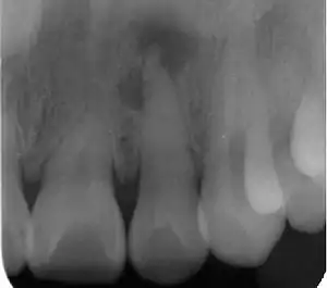
| Radiograph (X-ray) showing microdontia. Note also periapical lesion on the maxillary left lateral incisor. | |
| Specialty | Dentistry |
Teeth affected by microdontia may also have abnormal shape, and the abnormal size may affect the whole tooth, or only a part of the tooth.[1]
Definition
Males tend to have larger teeth than females,[1] and tooth size also varies by race.[1] Abnormal tooth size is defined by some as when the dimensions are more than 2 standard deviations from the average.[1] Microdontia is when the teeth are abnormally small, and macrodontia is when the teeth are abnormally large.
Classification
There are 3 types of microdontia:
True generalized
All the teeth are smaller than the normal size. True generalized microdontia is very rare, and occurs in pituitary dwarfism.[2][3] Due to decreased levels of growth hormone the teeth fail to develop to a normal size.[2]
Relative generalized
All the teeth are normal size but appear smaller relative to enlarged jaws.[3] Relative generalized microdontia may be the result of inheritance of a large jaw from one parent, and normal sized teeth from the other.[2]
Localized (focal)
Localized microdontia is also termed focal, or pseudo-microdontia. A single tooth is smaller than normal.[3] Localized microdontia is far more common than generalized microdontia,[2] and is often associated with hypodontia (reduced number of teeth).[1] The most commonly involved tooth in localized microdontia is the maxillary lateral incisor, which may also be shaped like an inverted cone (a "peg lateral").[3] Peg laterals typically occur on both sides,[2] and have short roots.[2] Inheritance may be involved,[2] and the frequency of microdontia in the upper laterals is just under 1%.[1] The second most commonly involved tooth is the maxillary third molars,[3] and after this supernumerary teeth.[3]
Causes
There are many potential factors involved.[4]
- Congenital hypopituitarism[1]
- Ectodermal dysplasia[1]
- Down syndrome[1]
- Ionizing radiation to the jaws during tooth development (odontogenesis)[1]
- Chemotherapy during tooth development[4]
- Marshall syndrome[4]
- Rieger syndrome[4]
- Focal dermal hypoplasia[4]
- Silver-Russell syndrome[4]
- Williams syndrome[4]
- Gorlin-Chaudhry-Moss syndrome[4]
- Coffin–Siris syndrome[4]
- Salamon syndrome[4]
- Cleft lip and palate[4]
Others include trichorhinopharyngeal, odontotrichomelic, neuroectodermal and dermo-odontodysplasia syndromes.[4]
Treatment
Unerupted microdonts may require surgical removal to prevent the formation of cysts.[2] Erupted microdonts, peg laterals especially, may cause cosmetic concern. Such teeth may be restored to resemble normal sized teeth,[2] typically with composite build ups or crowns.[4] Orthodontics may be required in severe cases to close gaps between the teeth.[4]
Epidemiology
Females are affected more than males,[4] and the condition occurs in permanent (adult) teeth more than deciduous (baby teeth or milk teeth).[4]
References
- Poulsen S; Koch G (2013). Pediatric dentistry: a clinical approach (2nd ed.). Chichester, UK: Wiley-Blackwell. p. 191. ISBN 9781118687192.
- Ibsen OAC; Phelan JA (2014). Oral Pathology for the Dental Hygienist (6th ed.). Elsevier Health Sciences. pp. 164–165. ISBN 9780323291309.
- Regezi JA; Scuibba JJ; Jordan RCK (2012). Oral pathology : clinical pathologic correlations (6th ed.). St. Louis, Mo.: Elsevier/Saunders. p. 373. ISBN 978-1-4557-0262-6.
- Laskaris G (2011). Color Atlas of Oral Diseases in Children and Adolescents. Thieme. p. 2. ISBN 9783131604712.