Iridocorneal endothelial syndrome
| Iridocorneal endothelial syndrome | |
|---|---|
 | |
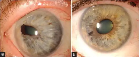
| a-b)Iridocorneal endothelial syndrome | |
Iridocorneal Endothelial (ICE) syndromes are a spectrum of diseases characterized by slowly progressive abnormalities of the corneal endothelium and features including corneal edema, iris distortion, and secondary angle-closure glaucoma. [1,2,4] ICE syndromes are predominantly unilateral and nonhereditary [1,2,4]. The condition occurs in predominantly middle-aged women [1,3,4].
Signs and Symptoms
Many cases are asymptomatic, however patients many have decreased vision, glare, monocular diplopia or polyopia, and noticeable iris changes [2,6]. On exam patients have normal to decreased visual acuity, and a “beaten metal appearance” of the corneal endothelium, corneal edema, increased intraocular pressure, peripheral anterior synechiae, and iris changes [1,2,6].
Mechanism
The exact mechanism is unknown, however there appears to be a component of abnormal corneal endothelium that proliferates onto the iris forming a membrane that then obstructs the trabecular meshwork, leading to iris distortion [1,2]. Nodule formation can also occur when the abnormal corneal endothelium causes contractions around the iris stroma [1]. Herpesvirus DNA has been identified in some patients following keratoplasty, suggesting the possibility that herpes simplex virus may induce the abnormal endotheliazation in the anterior chamber angle and on the surface of the iris [2,3,5].
Variations
The Chandler variant of ICE is characterized by pathology on the inner surface of the cornea leading to abnormal endothelial pump function [2,6]. Other features include possible mild iris changes, corneal edema, and normal to slight elevations in intraocular pressure [1,6].
Cogan-Reese variant is characterized by multiple pigmented iris nodules [2,6]. This variant is most commonly unilateral and seen in middle-aged females [2].
Diagnosis
The diagnosis for this condition entails a complete eye exam that would indicate-changes in iris, swelling of cornea, high intra ocular pressure [1]
Treatment
Penetrating karatoplasty and endothelial keratoplasty can be used as treatments for severe cases of ICE [2,8]. Because glaucoma and elevated intraocular pressure are often present in ICE patients, long term follow up may be needed to ensure adequate intraocular pressures are maintained [2,7]
Prognosis
The disease is chronic and often progresses slowly. Prognosis is generally poor when associated with glaucoma [1,2].
References
- ↑ "What Is Iridocorneal Endothelial Syndrome (ICE)?". American Academy of Ophthalmology. 19 October 2020. Archived from the original on 15 July 2021. Retrieved 16 December 2021.
- [1] Friedman NJ, Kaiser PK, Pineda R. (2009). The Massachusetts Eye and Ear Infirmary Illustrated Manual of Ophthalmology. Chapter 7: Iris and Pupils (pp. 285–287). Philadelphia PA:W.B. Saunders Company.
- [2] Weisenthal RW. 2012-2013 Basic and Clinical Science Course, Section 8, Chapter 12: External Disease and Cornea (pp 344–345). San Francisco CA: American Academy of Ophthalmology The Eye M.D. Association.
- [3] Alvardo JA, Underwood JL, Green WR, et al. (1994) Detection of herpes simplex viral DNA in the iridocorneal endothelial syndrome. Archives of Ophthalmology, 112(12), 1601-1609
- [4] Carpel EF. (2005). Iridocorneal endothelial syndrome. In: Krachmer JH, Mannis MJ, Holland EJ. Cornea. 2nd ed. Vol 1. Chapter 79 (pp 975–985). Philadelphia: Elsever/Mosby
- [5] Groh MJ, Seitz B, Schumacher S, Naumann GO. Detection of herpes simplex virus in aqueous humor in iridocorneal endothelial (ICE) syndrome. Cornea. 1999;18(3):359-360.
- [6] Herde J. Iridocorneal endothelial syndrome (ICE-S): classification, clinical picture, diagnosis. Klin Monatsbl Augenheilkd. 2005;222(10):797-801
- [7] Price MO, Price FW Jr. Descemet stripping with endothelial keratoplasty for treatment of iridocorneal endothelial syndrome. Cornea. 2007;26(4):493-497.
External links
| Classification |
|
|---|---|
| External resources |
|
- Facts About the Cornea and Corneal Disease The National Eye Institute (NEI).