Endophthalmitis
| Endophthalmitis | |
|---|---|
 | |
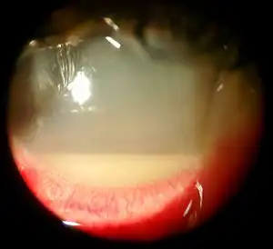
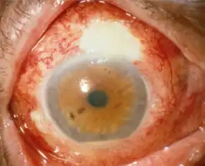
| A hazy eye with a hypopyon | |
| Specialty | Ophthalmology |
| Symptoms | Vision loss, eye pain, red eye, hypopyon, corneal edema[1] |
| Complications | Glaucoma, orbital cellulitis, loss of the eye[1] |
| Types | Exogenous, endogenous[1] |
| Causes | Bacteria (95%), fungi (5%)[1] |
| Risk factors | Eye surgery, eye injury, eye injections[1] |
| Diagnostic method | Eye examination, microbial culture of the eye[1] |
| Differential diagnosis | Toxic anterior segment syndrome, uveitis, vitreous hemorrhage[1] |
| Prevention | Povidone-iodine before eye surgery[2] |
| Treatment | Antibiotics, vitrectomy, corticosteroids, atropine[1] |
Endophthalmitis is inflammation of the interior cavity of the eye, usually caused by infection.[1] Symptoms may include vision loss, eye pain, red eye, hypopyon, and corneal edema.[1] Complications may include glaucoma, orbital cellulitis, loss of the eye itself.[1]
It usually is due to a bacterial infection (95%), though may also occur due to fungi (5%).[1] Risk factors include eye surgery, eye injury, and eye injections.[1] Occasionally it may spread from other areas of the body.[1] Diagnosis is based on eye examination and microbial culture of the eye.[1]
Treatment involves antibiotics, such as vancomycin and ceftazidime or amikacin, which are typically given by injection into the eye.[1][2] Amphotericin B or voriconazole may be used for fungal infections.[2] Other measures may include vitrectomy, corticosteroids, and atropine eye drops.[1]
Endophthalmitis, unrelated to eye procedures, is uncommon.[2] Following open globe injury it occurs in up to 15 to 30% of cases.[2] In the United States it occurs in about 4 per 10,000 cataract surgeries and 1 per 20,000 eye injections.[1][2]
Signs and symptoms

There is usually a history of recent eye surgery or penetrating trauma to the eye. Symptoms include severe pain, vision loss, and intense redness of the conjunctiva.[3] Hypopyon can be present and should be looked for on examination by a slit lamp. It can first present with the 'black dot sign' (Martin-Farina sign), where patients may report a small area of loss of vision that resembles a black dot or fly.
An eye exam should be considered in systemic candidiasis, as up to 3% of cases of candidal blood infections lead to endophthalmitis.
Complications
- Panophthalmitis — Progression to involve all the coats of the eye.[4]
- Corneal ulcer[4]
- Orbital cellulitis[4]
- Impairment of vision
- Complete loss of vision
- Loss of eye architecture
- Enucleation
Cause
- Bacteria: N. meningitidis, Staphylococcus aureus, S. epidermidis, S. pneumoniae, other streptococcal spp., Cutibacterium acnes, Pseudomonas aeruginosa, other gram negative organisms.[5]
- Viruses: Herpes simplex virus.[5]
- Fungi: Candida spp.[5] Fusarium
- Parasites: Toxoplasma gondii, Toxocara.[5]
The most common source of infectious following cataract surgery has been attributed to a contaminated intaocular solution (i.e. irrigation solution, viscoelastic, or diluted antibiotic), although there is a large diversity of microorganisms that can travel via various routes including the operating room environment, phacoemulsifcation machine, surgical instruments, topical anesthetics, intraocular lens, autoclave solution, and cotton wool swabs.[6]
Late-onset endophthalmitis is mostly caused by Cutibacterium acnes.[7]
Causative organisms are not present in all cases. Endophthalmitis can emerge by entirely sterile means, e.g. an allergic reaction to a drug administered intravitreally.
Diagnosis
Diagnosis: Microbiology testing PCR TASS vs Infectious endophthalmitis[3]
Classification
The following are within the classification of endophthalmitis:[8]
- Acute-onset postoperative endophthalmitis
- Delayed-onset postoperative endophthalmitis
- Bleb-associated endophthalmitis
- Postintravitreal injection endophthalmitis
- Posttraumatic endophthalmitis
- Endogenous endophthalmitis
Prevention
Preventative antibiotics (injections in the eye with cefuroxime) during cataract surgery lowers the chance of endophthalmitis.[9] Also, antibiotic eye drops (levofloxacin or chloramphenicol) with antibiotic injections (cefuroxime or penicillin) probably lowers the chance of endophthalmitis compared with injections or eye drops alone.[9] Separate studies from the research showed that a periocular injection of penicillin with chloramphenicol-suphadimidine eye drops,[10] and an intracameral cefuroxime injection with topical levofloxacin reduces endophthalmitis following cataract surgery.[11]
In the case of intravitreal injections, however, antibiotics are not effective. Studies have demonstrated no difference between rates of infection with and without antibiotics when intravitreal injections are performed.[12] The only consistent method of antibioprophylaxis in this instance is a solution of povidone-iodine applied pre-injection.[13]
Treatment
Treatment may involve intravitreal injections of antibiotics. Injections of vancomycin and ceftazidime are routine. Even though antibiotics can have negative impacts on the retina in high concentrations, visual acuity worsens in 65% of endophthalmitis and prognosis gets poorer the longer an infection goes untreated makes immediate intervention necessary.[14] Endophthalmitis may also require an urgent surgery (pars plana vitrectomy), and evisceration may be necessary to remove a severe and intractable infection which could result in a blind and painful eye.
Steroids may be injected intravitreally if the cause is allergic.
In acute endophthalmitis, it is unclear if adding steroids to antibiotics improves outcomes.[15]
References
- 1 2 3 4 5 6 7 8 9 10 11 12 13 14 15 16 17 18 Simakurthy, S; Tripathy, K (January 2022). "Endophthalmitis". PMID 32644505.
{{cite journal}}: Cite journal requires|journal=(help) - 1 2 3 4 5 6 Relhan, N; Forster, RK; Flynn HW, Jr (March 2018). "Endophthalmitis: Then and Now". American journal of ophthalmology. 187: xx–xxvii. doi:10.1016/j.ajo.2017.11.021. PMID 29217351.
- 1 2 "Endophthalmitis". The Lecturio Medical Concept Library. Archived from the original on 19 July 2021. Retrieved 19 July 2021.
- 1 2 3 Goldenberg DT, Harinandan A, Walsh MK, Hassan T (Spring 2010). "Serratia Marcescens Endophthalmitis After 20-Gauge Pars Plana Vitrectomy". Retinal Cases & Brief Reports. 4 (2): 140–2. doi:10.1097/ICB.0b013e31819955bf. PMID 25390387.
- 1 2 3 4 Forbes BA, Sahm DF, Weissfeld AS. Bailey & Scott's Diagnostic Microbiology. 12th Edition. Mosby Elsevier, 2007. p. 834.
- ↑ Park, Jeff; Popovic, Marko M.; Balas, Michael; El-Defrawy, Sherif R.; Alaei, Ravin; Kertes, Peter J. (January 2022). "Clinical features of endophthalmitis clusters after cataract surgery and practical recommendations to mitigate risk: systematic review". Journal of Cataract and Refractive Surgery. 48 (1): 100–112. doi:10.1097/j.jcrs.0000000000000756. ISSN 0886-3350. PMID 34538777. S2CID 237574618. Archived from the original on 2022-08-01. Retrieved 2022-07-24.
- ↑ Shirodkar AR, Pathengay A, Flynn HW, Albini TA, Berrocal AM, Davis JL, Lalwani GA, Murray TG, Smiddy WE, Miller D (March 2012). "Delayed- versus acute-onset endophthalmitis after cataract surgery". Am. J. Ophthalmol. 153 (3): 391–398.e2. doi:10.1016/j.ajo.2011.08.029. PMC 3381653. PMID 22030353.
- ↑ Vaziri, Kamyar; Schwartz, Stephen G.; Kishor, Krishna; Flynn, Harry W. (2015). "Endophthalmitis: state of the art". Clinical Ophthalmology (Auckland, N.Z.). 9: 95–108. doi:10.2147/OPTH.S76406. ISSN 1177-5467. Archived from the original on 27 November 2022. Retrieved 27 November 2022.
- 1 2 Gower EW, Lindsley K, Tulenko SE, Nanji AA, Leyngold I, McDonnell PJ (February 2017). "Perioperative antibiotics for prevention of acute endophthalmitis after cataract surgery". Cochrane Database Syst Rev. 2017 (2): CD006364. doi:10.1002/14651858.CD006364.pub3. PMC 5375161. PMID 28192644.
- ↑ Christy NE, Sommer A (1979). "Antibiotic prophylaxis of postoperative endophthalmitis". Annals of Ophthalmology. 11 (8): 1261–1265. PMID 318049.
- ↑ Endophthalmitis Study Group; European Society of Cataract; Refractive Surgeons (2007). "Prophylaxis of postoperative endophthalmitis following cataract surgery: results of the ESCRS multicenter study and identification of risk factors". Journal of Cataract and Refractive Surgery. 33 (6): 978–988. doi:10.1016/j.jcrs.2007.02.032. PMID 17531690. S2CID 37697458.
- ↑ d’Azy, Cédric Benoist; Pereira, Bruno; Naughton, Geraldine; Chiambaretta, Frédéric; Dutheil, Frédéric (2016-06-03). "Antibioprophylaxis in Prevention of Endophthalmitis in Intravitreal Injection: A Systematic Review and Meta-Analysis". PLOS ONE. 11 (6): e0156431. Bibcode:2016PLoSO..1156431B. doi:10.1371/journal.pone.0156431. ISSN 1932-6203. PMC 4892688. PMID 27257676.
- ↑ de Caro, John J.; Ta, Christopher N.; Ho, Hoai-Ky V.; Cabael, Lorella; Hu, Nan; Sanislo, Steven R.; Blumenkranz, Mark S.; Moshfeghi, Darius M.; Jack, Robert (2008-06-01). "Bacterial contamination of ocular surface and needles in patients undergoing intravitreal injections". Retina (Philadelphia, Pa.). 28 (6): 877–883. doi:10.1097/IAE.0b013e31816b3180. ISSN 0275-004X. PMID 18536606. S2CID 25819637.
- ↑ Dossarps, Denis; Bron, Alain M.; Koehrer, Philippe; Aho-Glélé, Ludwig S.; Creuzot-Garcher, Catherine (2016). "Endophthalmitis after intravitreal injections: Incidence, presentation, management, and visual outcome". American Journal of Ophthalmology. 160 (1): 17–25.e1. doi:10.1016/j.ajo.2015.04.013. PMID 25892127.
- ↑ Emami, Sara; Kitayama, Ken; Coleman, Anne L. (2022-06-06). "Adjunctive steroid therapy versus antibiotics alone for acute endophthalmitis after intraocular procedure". The Cochrane Database of Systematic Reviews. 6: CD012131. doi:10.1002/14651858.CD012131.pub3. ISSN 1469-493X. PMC 9169535. PMID 35665485. Archived from the original on 2022-07-24. Retrieved 2022-07-24.
External links
| Classification | |
|---|---|
| External resources |