First-degree atrioventricular block
| First-degree AV block | |
|---|---|
| Other names: First degree heart block, PR prolongation | |
 | |
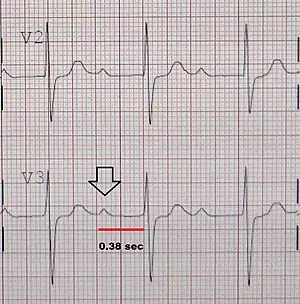
| An ECG showing a first degree AV block of greater than 300 ms | |
| Specialty | Cardiology |
| Symptoms | None[1] |
| Complications | Progression to second or third degree AV block[2] |
| Causes | Normal, heart attack, myocarditis, high blood potassium, certain medications[3] |
| Diagnostic method | Electrocardiogram (ECG)[3] |
| Differential diagnosis | Second-degree AV block, third-degree AV block[2] |
| Treatment | Routine follow-up, avoidance of certain medication[2] |
| Prognosis | Good[3] |
| Frequency | 5% of people over 50 years[4] |
First-degree atrioventricular block (first degree AV block) is were the electrical signal from the upper to lower chambers of the heart is delayed but not interrupted.[3] It generally results in no symptoms.[1] Rarely there may be shortness of breath, tiredness, and lightheadedness.[2] Complications may include atrial fibrillation or progress to second- or third-degree AV block.[2]
It occur occur normally in younger people and athletes.[1] Other causes include a heart attack, myocarditis, high blood potassium, and certain medications such as beta blockers, calcium channel blockers, and digoxin.[3] Diagnosis is by an electrocardiogram (ECG) showing a PR interval of greater than 200 milliseconds without any dropped beats.[3] The condition is considered "marked" is greater than 300 milliseconds.[3] It is a type of atrioventricular block.[3]
No specific treatment is generally required.[1] Medications that can worsen the condition may be stopped.[3] Rarely in those with marked disease and symptoms a pacemaker may be placed.[2] In young people brief episodes of first degree AV block and common though ongoing issues affects less than 1%.[4] In becomes more common with age affecting about 5% of people over the age of 50.[4] Males are more commonly affected than females.[2]
Signs and symptoms
It generally results in no symptoms.[1] Rarely there may be shortness of breath, tiredness, and lightheadedness.[2] Complications may include atrial fibrillation or progress to second- or third-degree AV block.[2]
Causes
The most common causes of first-degree heart block are an AV nodal disease, enhanced vagal tone (for example in athletes), myocarditis, acute myocardial infarction (especially acute inferior MI), electrolyte disturbances and medication. The medications that most commonly cause first-degree heart block are those that increase the refractory time of the AV node, thereby slowing AV conduction. These include calcium channel blockers, beta-blockers, cardiac glycosides, and anything that increases cholinergic activity such as cholinesterase inhibitors.
Diagnosis
In normal individuals, the AV node slows the conduction of electrical impulse through the heart. This is manifest on a surface electrocardiogram (ECG) as the PR interval. The normal PR interval is from 120 ms to 200 ms in length. This is measured from the initial deflection of the P wave to the beginning of the QRS complex.
In first-degree heart block, the diseased AV node conducts the electrical activity more slowly. This is seen as a PR interval greater than 200 ms in length on the surface ECG. It is usually an incidental finding on a routine ECG.
First-degree heart block does not require any particular investigations except for electrolyte and drug screens, especially if an overdose is suspected.
Treatment
The management includes identifying and correcting electrolyte imbalances and withholding any offending medications. This condition does not require admission unless there is an associated myocardial infarction. Even though it usually does not progress to higher forms of heart block, it may require outpatient follow-up and monitoring of the ECG, especially if there is a comorbid bundle branch block. If there is a need for treatment of an unrelated condition, care should be taken not to introduce any medication that may slow AV conduction. If this is not feasible, clinicians should be very cautious when introducing any drug that may slow conduction; and regular monitoring of the ECG is indicated.
Prognosis
Isolated first-degree heart block has no direct clinical consequences. There are no symptoms or signs associated with it. It was originally thought of as having a benign prognosis. In the Framingham Heart Study, however, the presence of a prolonged PR interval or first degree AV block doubled the risk of developing atrial fibrillation, tripled the risk of requiring an artificial pacemaker, and was associated with a small increase in mortality. This risk was proportional to the degree of PR prolongation.[5]
A subset of individuals with the triad of first-degree heart block, right bundle branch block, and either left anterior fascicular block or left posterior fascicular block (known as trifascicular block) may be at an increased risk of progression to complete heart block.
References
- 1 2 3 4 5 "Atrioventricular Block - Cardiovascular Disorders". Merck Manuals Professional Edition. Archived from the original on 6 April 2010. Retrieved 29 December 2020.
- 1 2 3 4 5 6 7 8 9 Oldroyd, SH; Quintanilla Rodriguez, BS; Makaryus, AN (January 2020). "First Degree Heart Block". PMID 28846254.
{{cite journal}}: Cite journal requires|journal=(help) - 1 2 3 4 5 6 7 8 9 Kashou, AH; Goyal, A; Nguyen, T; Chhabra, L (January 2020). "Atrioventricular Block". PMID 29083636.
{{cite journal}}: Cite journal requires|journal=(help) - 1 2 3 Saksena, Sanjeev; Camm, A. John (2011). Electrophysiological Disorders of the Heart E-Book: Expert Consult. Elsevier Health Sciences. p. 521. ISBN 978-1-4377-0971-1. Archived from the original on 2021-08-28. Retrieved 2020-12-29.
- ↑ Cheng S, Keyes MJ, Larson MG, McCabe EL, Newton-Cheh C, Levy D, Benjamin EJ, Vasan RS, Wang TJ (2009). "Long-term outcomes in individuals with prolonged PR interval or first-degree atrioventricular block". JAMA. 301 (24): 2571–2577. doi:10.1001/jama.2009.888. PMC 2765917. PMID 19549974.
External links
| Classification | |
|---|---|
| External resources |