Right bundle branch block
| Right bundle branch block | |
|---|---|
 | |
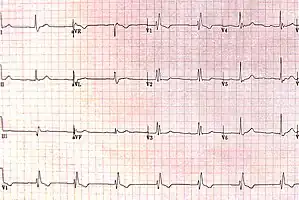
| ECG characteristics of a typical RBBB showing wide QRS complexes with a terminal R wave in lead V1 and a prolonged S wave in lead V6. | |
| Specialty | Cardiology |
| Symptoms | None[1] |
| Complications | None[1] |
| Causes | Unknown, heart damage, increased right ventricular pressure, high blood potassium[1] |
| Diagnostic method | Electrocardiogram (ECG)[1] |
| Differential diagnosis | Left bundle branch block, ventricular tachycardia, Brugada syndrome[1] |
| Treatment | Based on underlying disorder[1] |
| Frequency | Fairly common[2] |
Right bundle branch block (RBBB) is a type of electrical conduction abnormality of the heart seen on the electrocardiogram (ECG).[1] In and of itself, it results in no symptoms and generally results in no complications.[1] It may be temporary or permanently present.[2]
Causes include heart damage, such as a heart attack or myocarditis, increased right ventricular pressure, such as with pulmonary embolism or cor pulmonale, and rarely high blood potassium.[1] Though some cases occurs without any specific cause.[2] The underlying mechanism involves damage to the right bundle branch.[1]
Diagnosis requires that the QRS complex is greater than 120 ms and an rsR' wave is present in lead V1 or V2.[2] The T waves are generally flipped in V1 and V2.[2] When the QRS duration is less than 120 ms, but the other criteria are present, it is called an incomplete RBBB.[1] Its presence does not interfere with the diagnosis of a heart attack.[1]
No specific treatment is generally required.[1] In the setting of heart failure, cardiac resynchronization therapy may be indicated.[1] RBBB is present in about 0.2% to 0.8% of ECGs.[2] Up to 11% of people are affected by the age of 80.[1]
Signs and symptoms
In and of itself, it results in no symptoms.[1]
Causes
Common causes are Normal variant, right ventricular hypertrophy or strain, cCongenital heart disease such as atrial septal defect and Ischemic heart disease.[3] In addition, a right bundle branch block may also result from Brugada syndrome, pulmonary embolism, rheumatic heart disease, myocarditis, cardiomyopathy, or hypertension.
Diagnosis
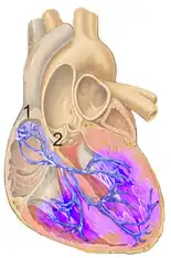
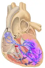
The criteria to diagnose a right bundle branch block on the electrocardiogram:
- The heart rhythm must originate above the ventricles (i.e., sinoatrial node, atria or atrioventricular node) to activate the conduction system at the correct point.
- The QRS duration must be more than 100 ms (incomplete block) or more than 120 ms (complete block).[4]
- There should be a terminal R wave in lead V1 (often called "R prime," and denoted by R, rR', rsR', rSR', or qR).
- There must be a prolonged S wave in leads I and V6 (sometimes referred to as a "slurred" S wave).
The T wave should be deflected opposite the terminal deflection of the QRS complex. This is known as appropriate T wave discordance with bundle branch block. A concordant T wave may suggest ischemia or myocardial infarction.
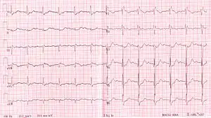 RBBB with associated first degree AV block
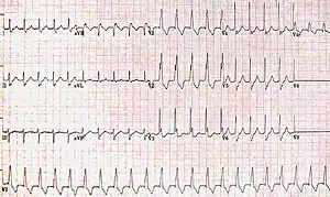 RBBB with associated tachycardia
 RBBB
RBBB
Treatment
The underlying condition may be treated by medications to control hypertension or diabetes, if they are the primary underlying cause. If coronary arteries are blocked, an invasive coronary angioplasty may relieve the impending RBBB.[5]
Epidemiology
RBBB is present in about 0.2% to 0.8% of ECGs.[2] Up to 11% of people are affected by the age of 80.[1]
See also
References
- 1 2 3 4 5 6 7 8 9 10 11 12 13 14 15 16 17 Harkness, WT; Hicks, M (January 2020). "Right Bundle Branch Block". PMID 29939649.
{{cite journal}}: Cite journal requires|journal=(help) - 1 2 3 4 5 6 7 Roberts, David (2019). Mastering the 12-Lead EKG. Springer Publishing Company. p. 316. ISBN 978-0-8261-8194-7. Archived from the original on 2021-08-29. Retrieved 2020-12-30.
- ↑ Goldman, Lee (2011). Goldman's Cecil Medicine (24th ed.). Philadelphia: Elsevier Saunders. pp. 400–401. ISBN 978-1437727883.
- ↑ "Lesson VI - ECG Conduction Abnormalities". Archived from the original on 2009-01-16. Retrieved 2009-01-07.
- ↑ "Right Bundle Branch Block". www.symptoma.com. Archived from the original on 2017-10-05. Retrieved 2015-08-13.
External links
| Classification | |
|---|---|
| External resources |