Atrial tachycardia
| Atrial tachycardia | |
|---|---|
 | |
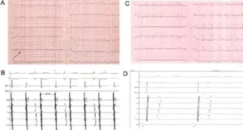
| a) Atrial tachycardia with 2:1 conduction is detectable; the atrial waves morphology involves the likely diagnosis of left sided atrial tachycardia b) shows left atrial tachycardia with 2:1 conduction to the ventricles; the distal dipole of the catheter positioned inside the coronary sinus is the first one to be recording atrial signals c)post-procedural surface ECG sinus rhythm is visible d) intracardiac electrograms confirm the earliest atrial activation to be at the level of the high right atrium | |
| Specialty | Cardiology |
Atrial tachycardia is a type of heart rhythm problem in which the heart's electrical impulse comes from an ectopic pacemaker (that is, an abnormally located cardiac pacemaker) in the upper chambers (atria) of the heart, rather than from the sinoatrial node, the normal origin of the heart's electrical activity. As with any other form of tachycardia (rapid heart beat), the underlying mechanism can be either the rapid discharge of an abnormal focus, the presence of a ring of cardiac tissue that gives rise to a circle movement (reentry),[1] or a triggered rapid rhythm due to other pathological circumstances (as would be the case with some drug toxicities, such as digoxin toxicity).
Classification
Forms of atrial tachycardia (ATach) include multifocal atrial tachycardia (MAT), focal atrial tachycardia and atrial flutter.[2] Paroxysmal atrial tachycardia (PAT) is an episode of arrhythmia that begins and ends abruptly.[3]
Cause
Atrial tachycardia tends to occur in individuals with structural heart disease, with or without heart failure, and ischemic coronary artery disease. However, focal atrial tachycardia often occurs in healthy individuals without structural heart disease. Other possible etiologies are listed below:[2]
- Hypoxia
- Pulmonary disease
- Ischemic heart disease
- Stimulants: cocaine, caffeine, chocolate, ephedra
- Alcohol
- Metabolic disturbances
- Digoxin toxicity
- Heightened sympathetic tone
A study noted 10 to 15% of patients presenting for supraventricular tachycardia ablation had atrial tachycardia.[2]
Diagnosis
Electrocardiographic features include:[2]
- Atrial rate: 100 to 250 BPM
- Ventricular conduction can be variable
- Irregular or irregularly irregular in the setting of variable AV block
- Regular if 1 to 1, 2 to 1, or 4 to 1 AV block
- P wave morphology
- Unifocal, but similar in morphology to each other
- Might be inverted
- Differs from normal sinus P wave
- May exhibit either long RP or short PR intervals
- Rhythm may be paroxysmal or sustained
- May demonstrate an increase in the rate at initiation (e.g., "warm up," or "rev up")
- May demonstrate a decrease in the rate at termination (e.g., "cool down")
Treatment
Initial management of focal atrial tachycardia should focus on addressing underlying causes: treating acute illness, cessation of stimulants, stress reduction, appropriately managing digoxin toxicity, or chronic disease management. The ventricular rate is controllable with the use of beta blockers or calcium channel blockers. If atrial tachyarrhythmia persists and the patient is symptomatic, the patient may benefit from class IA, IC, or class III antiarrhythmics. Catheter ablation of focal atrial tachycardia may be appropriate in patients failing medical therapy.[2]
Epidemiology
A European study of young males applying for pilot licenses demonstrated that 0.34% had asymptomatic atrial tachycardia and 0.46% had symptomatic atrial tachycardia.[2][4]
References
- ↑ Curr Opin Cardiol. 2001 Jan;16(1):1–7. "Basic mechanisms of reentrant arrhythmias". Antzelevitch C.
- 1 2 3 4 5 6 Mark Liwanag, Cameron Willoughby (2020). "Atrial Tachycardia". StatPearls. PMID 31194392. Archived from the original on 2022-10-23. Retrieved 2022-10-18.
{{cite journal}}: CS1 maint: uses authors parameter (link) Text was copied from this source, which is available under a Creative Commons Attribution 4.0 International License Archived 2017-10-16 at the Wayback Machine.
 - ↑ "Understanding Paroxysmal Atrial Tachycardia (PAT)". Healthline. June 12, 2012. Archived from the original on April 26, 2022. Retrieved October 18, 2022.
- ↑ Poutiainen, A M (1999). "Prevalence and natural course of ectopic atrial tachycardia". European Heart Journal. 20 (9): 694–700. doi:10.1053/euhj.1998.1313. PMID 10208790.
External links
| Classification | |
|---|---|
| External resources |