Subconjunctival bleeding
| Subconjunctival bleeding | |
|---|---|
| Other names: Subconjunctival hemorrhage, subconjunctival haemorrhage, hyposphagma | |
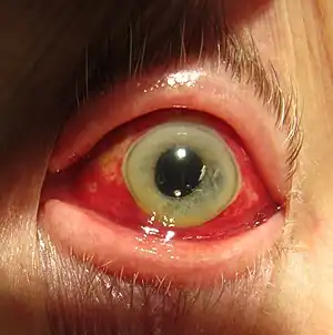 | |
| Subconjunctival hemorrhage resulting in red coloration of the white of the eye. | |
| Specialty | Ophthalmology |
| Symptoms | Red spot over whites of the eye, little to no pain[1] |
| Complications | None[2] |
| Duration | Two to three weeks[2] |
| Types | Traumatic, spontaneous[2] |
| Causes | Coughing, vomiting, direct injury[2] |
| Risk factors | High blood pressure, diabetes, older age[2] |
| Diagnostic method | Based on the appearance[2] |
| Differential diagnosis | Open globe, retrobulbar hematoma, conjunctivitis, pterygium[2] |
| Treatment | No specific treatment[3] |
| Medication | Artificial tears[2] |
| Prognosis | Good, 10% risk of reoccurance[2] |
| Frequency | Common[4] |
Subconjunctival bleeding, also known as subconjunctival hemorrhage, is bleeding from a small blood vessel over the whites of the eye.[1] It results in a red spot in the white of the eye.[1] There is generally little to no pain and vision is not affected.[2][3] Typically only one eye is affected.[2]
Causes can include coughing, vomiting, heavy lifting, and direct injury including that from wearing contact lenses.[2] Risk factors include high blood pressure, diabetes, older age, blood thinners, and trauma including that from wearing contact lenses.[2] They occur in about 2% of newborns following a vaginal delivery.[2] The blood occurs between the conjunctiva and the episclera.[2] Diagnosis is largely based on the appearance.[2]
Usually no specific treatment is required and the condition improves in two to three weeks.[2] Artificial tears may be used to help with any irritation.[2] They occur relatively commonly.[4] Both sexes are affected equally.[2] Spontaneous bleeding occurs more commonly over the age of 50 while the traumatic type occurs more often in young males.[2]
Signs and symptoms
Subconjunctival bleeding is bleeding from a small blood vessel over the whites of the eye.[1] It results in one or more red spots in the white of the eye, usually noticed when looking in the mirror.[1] There is generally little to no pain and vision is not affected.[2][3] Typically only one eye is affected.[2]
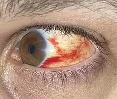
A subconjunctival bleeding initially appears bright-red underneath the transparent conjunctiva. Later, the bleeding may spread and become green or yellow as the hemoglobin is metabolized. It usually disappears within 2 weeks.[5]
 Minor subconjunctival bleed
.jpg.webp) After one week
After one week.jpg.webp) Same as prior after four weeks
Same as prior after four weeks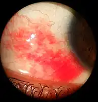 As viewed through a slit lamp
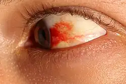 After 48 hours
Causes
Subconjunctival bleeding can occur without a trigger, or due to trauma, infection or bleeding disorder.[6] Mostly it occurs spontaneously, particularly in older people with more fragile blood vessels.[6]
It can be caused by head injury and trauma to the eye, or after eye surgery such as LASIK.[6][7] Other traumatic causes may arise from straining such as heavy lifting or vomiting, or from increased pressure in the chest and abdomen such as from being squeezed in a crowd.[6] Choking, or coughing may cause subconjunctival bleeding.[2] Another cause may be constipation.[2] Zygoma fracture results in lateral subconjunctival bleeding. Another cause is mask squeeze from diving and not equalizing mask pressure during descent.[8] Causes include atmospheric pressure changes such as those from diving deeply in water and aircraft altitude changes.[9]
Infections such as conjunctivitis can result in a subconjunctival bleed.[6] Other infections included Ebola, acute hemorrhagic conjunctivitis (caused by Enterovirus 70 or Coxsackie A virus), Leptospirosis.
Rarely there may be a serious cause such as a bleeding disorder or leukaemia, conditions in which the subconjunctival bleeding may be recurrent.[6]
Subconjunctival bleeding in children may be associated with scurvy (vitamin C deficiency), whooping cough, malaria, purpura,[6][10] abuse or traumatic asphyxia syndrome.[11]
Risk factors
Risk factors include high blood pressure, diabetes, older age, blood thinners, and wearing contact lenses.[2]
Diagnosis
Diagnosis is based on the appearance, by noting the typical finding of bright red discoloration confined to the white portion (sclera) of the eye.[2]
Management
Subconjunctival bleeding is typically a self-limiting condition that requires no specific treatment unless there is evidence of an infection or there has been significant injury. Artificial tears may be applied four to six times a day if the eye feels dry or scratchy.[5][6] The elective use of aspirin is typically discouraged.[6]
References
- 1 2 3 4 5 Boyd, Kierstan (23 April 2020). "What is a Subconjunctival Hemorrhage?". American Academy of Ophthalmology. Archived from the original on 11 April 2021. Retrieved 11 April 2021.
- 1 2 3 4 5 6 7 8 9 10 11 12 13 14 15 16 17 18 19 20 21 22 23 24 25 26 Doshi, R; Noohani, T (January 2020). "Subconjunctival Hemorrhage". StatPearls. PMID 31869130. Archived from the original on 2021-08-29. Retrieved 2021-04-12.
- 1 2 3 Cronau, H; Kankanala, RR; Mauger, T (15 January 2010). "Diagnosis and management of red eye in primary care". American Family Physician. 81 (2): 137–44. PMID 20082509.
- 1 2 Gold, Daniel H.; Lewis, Richard Alan (2010). Clinical Eye Atlas. Oxford University Press. p. 82. ISBN 978-0-19-534217-8. Archived from the original on 2021-08-29. Retrieved 2020-05-07.
- 1 2 Graham, Robert H. (11 June 2019). "Red Eye: Background, Pathophysiology and Etiology, Epidemiology and Prognosis". emedicine.medscape.com. Archived from the original on 11 April 2021. Retrieved 11 April 2021.
- 1 2 3 4 5 6 7 8 9 Sihota, Ramanjit; Tandon, Radhika (2019). "7. Diseases of the conjunctiva". Parsons' Diseases of the Eye (23rd ed.). Elsevier. p. 142. ISBN 978-81-312-5415-8. Archived from the original on 2021-08-29. Retrieved 2021-04-11.
- ↑ Vajpayee, R B; Balasubramanya, R; Rani, A; Sharma, N; Titiyal, J S; Pandey, R M (June 2003). "Visual performance after interface haemorrhage during laser in situ keratomileusis". The British Journal of Ophthalmology. 87 (6): 717–719. ISSN 0007-1161. PMID 12770968. Archived from the original on 2021-08-29. Retrieved 2021-04-12.
- ↑ Bowman, John C.; Gossman, William (4 June 2020). "Diving Mask Squeeze". StatPearls. StatPearls Publishing. Archived from the original on 29 August 2021. Retrieved 12 April 2021.
- ↑ Lynch, James H.; Deaton, Travis G. (2014). "Barotrauma With Extreme Pressures in Sport: From Scuba to Skydiving". Current Sports Medicine Reports. 13 (2): 107–112. doi:10.1249/JSR.0000000000000039. ISSN 1537-8918. Archived from the original on 2019-07-26. Retrieved 2021-04-12.
- ↑ Rothschild, Bruce M. (19 February 2021). "Scurvy Imaging: Practice Essentials, Radiography". Archived from the original on 12 April 2021. Retrieved 12 April 2021.
- ↑ Spitzer S. G; Luorno J.; Noël L. P. (2005). "Isolated subconjunctival hemorrhages in nonaccidental trauma". Journal of American Association for Pediatric Ophthalmology and Strabismus. 9 (1): 53–56. doi:10.1016/j.jaapos.2004.10.003. PMID 15729281.
External links
| Classification | |
|---|---|
| External resources |