Prostaglandin F receptor
Prostaglandin F receptor (FP) is a receptor belonging to the prostaglandin (PG) group of receptors. FP binds to and mediates the biological actions of Prostaglandin F2α (PGF2α). It is encoded in humans by the PTGFR gene.[5]
Gene
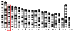
The PTGFR gene is located on human chromosome 1 at position p31.1 (i.e. 1p31.1), contains 7 exons, and codes for a G protein coupled receptor (GPCR) of the rhodopsin-like receptor family, Subfamily A14 (see rhodopsin-like receptors#Subfamily A14). PTGFR is expressed as two alternatively spliced transcript variants encoding different isoforms, FPA and FPB, which have different C-terminal lengths.[5][6][7] MicroRNA miR-590-3p binds to the Three prime untranslated region of the FP gene to repress its translation. miR-590-3p thus appears to be a negative regulator of FP expression in various cell types.[8]
Expression
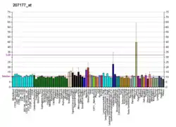
In humans, FP mRNA and/or protein is highly expressed in the uterine myometrium; throughout the eye (endothelium and smooth muscle cells of blood vessels of the iris), ciliary body and choroid plexus; ciliary muscle (circular muscle, collagenous connective tissues; sclera; and ovarian (follicles and corpus luteum). Studies in mice indicate that FP mRNA and/or protein is expressed in diverse tissues including the kidney (distal tubules), uterus, and ovary (Luteal cells of corpus luteum.[9][10]
Ligands
Activating ligands
The FP receptor is the least selective of the prostenoid receptors in that it is responsive to PGD2 and to a lesser extent PGE2 at concentrations close to those of PGF2α. Standard prostanoids have the following relative efficacies as receptor ligands in binding to and activating FP: PGF2α>PGD2>PGE2>PGI2=TXA2. In typical binding studies, PGF2α has one-half maximal binding and cell stimulating actions at ~1 nanomolar whereas PGD2 and PGE2 are ~5- to 10-fold and 10-100-fold weaker than this. The synthetic analogs that like PGF2α act as selective receptor agonists of FP viz., cloprostenol, flupostenol, latanoprost, and tafluprost (acid form) have FP binding affinities and stimulating potencies similar to PGF2α while others as enprostil, sulprostone, U46619, carbacyclin, and iloprost are considerably weaker FP agonists. Fluprostenol is a widely used clinically as a selective FP receptor agonist; latanoprost is a suitable substitute.[9]
Inhibiting ligands
Currently, there are no selective receptor antagonists for FP.[9]
Mechanism of cell activation
FP is classified as a contractile type of prostenoid receptor based on its ability, upon activation, to contract certain smooth muscle preparations and smooth muscle-containing tissues such as those of the uterus. When bound to PGF2α or other of its agonists, FP mobilizes primarily G proteins containing the Gq alpha subunit bound to of the Gq-Gβγ complex(i.e. Gqβγ). Gqβγ then dissociate into its Gq and Gβγ components which act to regulate cell signaling pathways. In particular, Gq stimulates cell signal pathways involving a) phospholipase C/IP3/cell Ca2+ mobilization/diacylglycerol/protein kinase Cs; calmodulin-modulated myosin light chain kinase; RAF/MEK/Mitogen-activated protein kinases; PKC/Ca2+/Calcineurin/Nuclear factor of activated T-cells; and the EGF cellular receptor.[7][11] In certain cells, activation of FP also stimulates G12/G13-Gβγ G proteins to activate the Rho family of GTPases signaling proteins and Gi-Gβγ G proteins to activateRaf/MEK/mitogen-activated kinase pathways.[11]
Functions
Studies using animals genetically engineered to lack FP and examining the actions of EP4 receptor agonists in animals as well as animal and human tissues indicate that this receptor serves various functions. It has been regarded as the most successful therapeutic target among the 9 prostanoid receptors.[11]
Eye
Animal and human studies have found that the stimulation of FP receptors located on Ciliary muscle and trabecular meshwork cells of the eye widens the drainage channels (termed the uveoscleral pathway) that they form. This increases the outflow of aqueous humor from the anterior chamber of the eye through Schlemm's canal to outside of the eyeball. The increase in aqueous humor outflow triggered by FP receptor activation reduces Intraocular pressure and underlies the widespread usage of FP receptor agonists to treat glaucoma. László Z. Bitó is credited with making critical studies to define this intraocular pressure-relieving pathway.[12] Three FP receptor agonists are approved for clinical use in the USA viz., travoprost, latanoprost, and bimatoprost, and two additional agonists are prescribed in Europe and Asia viz., unoprostone and tafluprost.[13]
Hair growth
Since FP receptors are expresses in human dermal papillae and the use of FP agonists to treat glaucoma has as a side-effect an increase in eyelash growth, it has been suggested that FP agonists may be useful for treating baldness. This is supported by studies in the stump-tailed Macaque primate model of androgen-induced scalp alopecia which have found that the FP agonist, latanoprost, promotes scalp hair growth. These studies have not yet been translated into baldness therapy in humans.[12]
Reproduction
FP receptor activation contributes to the regression of the corpus luteum and thereby the estrus cycle in many species of farm animals. However, it does not make these contributions in mice and its contribution to these functions in humans is controversial. The receptor has been in use as a target for decades to regulate the estrus cycle as well as to induce labor in pregnant farm animals[14][15] FP gene knockout in female mice blocks parturition. That is, these FP-/- mice fail to enter labor even if induced by oxytocin due to a failure in copus luteum regression and consequential failure to stop secreting progesterone (declining progesterone levels trigger labor).[14][15][16] Studies with monkey and human tissues allow that FP receptors may have a similar function in humans.[10]
Skin pigmentation
One side effect of applying FP receptor agonists to eyelashes in humans is the development of hyperpigmentation at nearby skin sites. Follow-up studies of this side effect indicated than human skin pigment-forming melanocyte cells express FP receptors and respond to FP receptor agonists by increasing their dendricites (projections to other cells) as well as to increase their tyrosinase activity. Since skin melanocytes use their dendrites to transfer the skin pigment melanin to skin keratinocytes thereby darkening skin and since tyrosinase is the rate-limiting enzyme in the synthesis of melanin, these studies suggest that FP receptor activation may be a useful means to increase skin pigmentation.[17]
Bone
PGF2α triggers the NFATC2 pathway stimulating skeletal muscle cell growth.[18] PGF2α, shown or presumed to operate by activating FP receptors, has complex effects on bone osteoclasts and osteoblasts to regulate Bone remodeling. However, further studies on the impact of the PGF2α-FP axis on bone are needed to better understand the pathophysiology underlying bone turnover and to identify this axis as a novel pharmacological target for the treatment of bone disorders and diseases.[12][19]
Inflammation and allergy
Unlike other prostaglandin receptors which have been shown in numerous studies to contribute to inflammatory and allergic responses in animal models, there are few studies on the function of FP receptors in these responses. Gene knockout studies in mice clearly show that FP mediates the late phase (thromboxane receptor mediates the early phase) of the tachycardia response to the pro-inflammatory agent, lipopolysaccharide.[16][20] PTGFR knockout mice also show a reduction in the development of pulmonary fibrosis normally caused by microbial invasion or bleomycin treatment. Finally, administration of PGF2α to mice causes an acute inflammatory response and elevated biosynthesis of PGF2α has been found in the tissues of patients with rheumatoid arthritis, psoriatic arthritis, and other forms of arthritis. While much further work is needed, these studies indicate that PGF2α-FP axis has some pro-inflammatory and anti-inflammatory effects in animals that may translate to humans.[7] The axis may likewise play role in human allergic responses: PGF2α causes airway constriction in normal and asthmatic humans and its presence in human sputum is related to sputum eosinophil levels.[21]
Cardiovascular system
PGF2α simulates an increase in systolic blood pressure in wild type but not FP(−/−) mice. Furthermore, FP(-/-) mice have significantly lower blood pressure, lower plasma renin levels, and lower plasma angiotensin-1 levels than wild-type mice, and FP agonists have a negative inotropic effect to weaken the strength of heart beating in rats. Finally, FP(−/−) mice deficient in the LDL receptor exhibit significantly less atherosclerosis than FP(+/+) LDL receptor-deficient mice. Activation of FP thus has pathophysiological consequences for the cardiovascular system relative to blood pressure, cardiac function, and atherosclerosis in animal models. The mechanism behind these FP effects and their relevancy to humans have not been elucidated.[12]
Clinical significance
Glaucoma
FP receptor agonists, specifically latanoprost, travoprost, bimatoprost, and tafluprost, are currently used as first-line drugs to treat glaucoma and other causes of intra-ocular hypertension (see Glaucoma#Medication).[22]
Hair growth
The FP receptor agonist, bimatoprost, in the form of an 0.03% ophthalmic solution termed Latisse, is approved by the US Food and Drug Administration to treat hypotrichosis of the eyelashes, in particular to darken and lengthen eyelashes for cosmetic purposes. Eyelid hypotrichosis caused by[17]
Veterinary uses
FP receptor agonists are used as highly effective agents to synchronize the oestrus cycles of farm animals and thereby to facilitate animal husbandry.[23]
Hair growth
Eyelash hypotrichosis due to the autoimmune disease, Alopecia areata, or to chemotherapy have been successfully treated with FP agonists in small Translational research studies. In a randomized, double-blind, placebo-controlled pilot study of 16 men with male pattern baldness (also termed androgenetic alopecia) topical application of the FP agonist, latanoprost, for 24 weeks produced a significant increase in scalp hair density. Despite these findings, however, a case report of one woman with female pattern hair loss found that injection of FP agonist bimatoprost failed to influence hair growth.[17]
Skin pigmentation
In preliminary studies, 3 Korean patients with periorbital vitiligo (i.e. skin blanching) were treated topically with the FP receptor agonist, latanoprost, for two months; the three patients experienced 20%, 50%, and >90% re-pigmentation of their vitiligo lesions. Fourteen patients with hypopigmented in their scarreed tissues were treated with the FP receptor agonist, bimatoprost, applied topically plus laser therapy and topical tretinoin or pimecrolimus. Most patients demonstrated significant improvement in their hypopigmentation, but the isolated effect of topical bimatoprost was not evaluated. These studies allow that FP receptor agonists may be useful for treating hypopigmentation such as occurs in scar tissue as well as diseases like vitiligo, tinea versicolor, and pityriasis alba.[17]
Genomic studies
The single-nucleotide polymorphism (SNP) A/G variant, rs12731181, located in the Three prime untranslated region of PTGFR has been associated with increased risk for hypertension in individuals from southern Germany; while this association was not replicated in other European populations, it was found in a Korean population. This SNP variant reduces the binging of MicroRNA miR-590-3p to PTGFR; since this binding represses translation of this gene, the rs127231181 variant acts to increase expression of the FP receptor.[8] PTGFR SNP variants rs6686438 and rs10786455s were associated with positive and SNP variants rs3753380, rs6672484, and rs11578155 in PTGFR were associated with negative responses to latanoprost for the treatment of Open-Angle Glaucoma in a Spanish population.[24] PTGFR SNP variants rs3753380 and rs3766355 were associated with a reduce response to latanoprost in a Chinese population study.[25]
References
- GRCh38: Ensembl release 89: ENSG00000122420 - Ensembl, May 2017
- GRCm38: Ensembl release 89: ENSMUSG00000028036 - Ensembl, May 2017
- "Human PubMed Reference:". National Center for Biotechnology Information, U.S. National Library of Medicine.
- "Mouse PubMed Reference:". National Center for Biotechnology Information, U.S. National Library of Medicine.
- "PTGFR prostaglandin F receptor [Homo sapiens (Human)] - Gene - NCBI".
- Zhang J, Gong Y, Yu Y (2010). "PG F(2α) Receptor: A Promising Therapeutic Target for Cardiovascular Disease". Frontiers in Pharmacology. 1: 116. doi:10.3389/fphar.2010.00116. PMC 3095374. PMID 21607067.
- Ricciotti E, FitzGerald GA (2011). "Prostaglandins and inflammation". Arteriosclerosis, Thrombosis, and Vascular Biology. 31 (5): 986–1000. doi:10.1161/ATVBAHA.110.207449. PMC 3081099. PMID 21508345.
- Xiao B, Gu SM, Li MJ, Li J, Tao B, Wang Y, Wang Y, Zuo S, Shen Y, Yu Y, Chen D, Chen G, Kong D, Tang J, Liu Q, Chen DR, Liu Y, Alberti S, Dovizio M, Landolfi R, Mucci L, Miao PZ, Gao P, Zhu DL, Wang J, Li B, Patrignani P, Yu Y (2015). "Rare SNP rs12731181 in the miR-590-3p Target Site of the Prostaglandin F2α Receptor Gene Confers Risk for Essential Hypertension in the Han Chinese Population". Arteriosclerosis, Thrombosis, and Vascular Biology. 35 (7): 1687–95. doi:10.1161/ATVBAHA.115.305445. PMID 25977569.
- "FP receptor - Prostanoid receptors - IUPHAR/BPS Guide to PHARMACOLOGY". www.guidetopharmacology.org.
- Kim SO, Markosyan N, Pepe GJ, Duffy DM (2015). "Estrogen promotes luteolysis by redistributing prostaglandin F2α receptors within primate luteal cells". Reproduction. 149 (5): 453–64. doi:10.1530/REP-14-0412. PMC 4380810. PMID 25687410.
- Moreno JJ (2017). "Eicosanoid receptors: Targets for the treatment of disrupted intestinal epithelial homeostasis". European Journal of Pharmacology. 796: 7–19. doi:10.1016/j.ejphar.2016.12.004. PMID 27940058. S2CID 1513449.
- Woodward DF, Jones RL, Narumiya S (2011). "International Union of Basic and Clinical Pharmacology. LXXXIII: classification of prostanoid receptors, updating 15 years of progress". Pharmacological Reviews. 63 (3): 471–538. doi:10.1124/pr.110.003517. PMID 21752876.
- Toris CB, Gulati V (2011). "The biology, pathology and therapeutic use of prostaglandins in the eye". Clinical Lipidology. 6 (5): 577–591. doi:10.2217/clp.11.42. S2CID 71994913.
- Ushikubi F, Sugimoto Y, Ichikawa A, Narumiya S (2000). "Roles of prostanoids revealed from studies using mice lacking specific prostanoid receptors". Japanese Journal of Pharmacology. 83 (4): 279–85. doi:10.1254/jjp.83.279. PMID 11001172.
- Sugimoto Y, Inazumi T, Tsuchiya S (2015). "Roles of prostaglandin receptors in female reproduction". Journal of Biochemistry. 157 (2): 73–80. doi:10.1093/jb/mvu081. PMID 25480981.
- Matsuoka T, Narumiya S (2008). "The roles of prostanoids in infection and sickness behaviors". Journal of Infection and Chemotherapy. 14 (4): 270–8. doi:10.1007/s10156-008-0622-3. PMID 18709530. S2CID 207058745.
- Choi YM, Diehl J, Levins PC (2015). "Promising alternative clinical uses of prostaglandin F2α analogs: beyond the eyelashes". Journal of the American Academy of Dermatology. 72 (4): 712–6. doi:10.1016/j.jaad.2014.10.012. PMID 25601618.
- Horsley V, Pavlath GK (2003). "Prostaglandin F2(alpha) stimulates growth of skeletal muscle cells via an NFATC2-dependent pathway". J Cell Biol. 161 (1): 111–8. doi:10.1083/jcb.200208085. PMC 2172881. PMID 12695501.
- Agas D, Marchetti L, Hurley MM, Sabbieti MG (2013). "Prostaglandin F2α: a bone remodeling mediator". Journal of Cellular Physiology. 228 (1): 25–9. doi:10.1002/jcp.24117. PMID 22585670. S2CID 206051942.
- Matsuoka T, Narumiya S (2007). "Prostaglandin receptor signaling in disease". TheScientificWorldJournal. 7: 1329–47. doi:10.1100/tsw.2007.182. PMC 5901339. PMID 17767353.
- Claar D, Hartert TV, Peebles RS (2015). "The role of prostaglandins in allergic lung inflammation and asthma". Expert Review of Respiratory Medicine. 9 (1): 55–72. doi:10.1586/17476348.2015.992783. PMC 4380345. PMID 25541289.
- Dams I, Wasyluk J, Prost M, Kutner A (2013). "Therapeutic uses of prostaglandin F(2α) analogues in ocular disease and novel synthetic strategies". Prostaglandins & Other Lipid Mediators. 104–105: 109–21. doi:10.1016/j.prostaglandins.2013.01.001. PMID 23353557.
- Coleman RA, Smith WL, Narumiya S (1994). "International Union of Pharmacology classification of prostanoid receptors: properties, distribution, and structure of the receptors and their subtypes". Pharmacological Reviews. 46 (2): 205–29. PMID 7938166.
- Ussa F, Fernandez I, Brion M, Carracedo A, Blazquez F, Garcia MT, Sanchez-Jara A, De Juan-Marcos L, Jimenez-Carmona S, Juberias JR, Martinez-de-la-Casa JM, Pastor JC (2015). "Association between SNPs of Metalloproteinases and Prostaglandin F2α Receptor Genes and Latanoprost Response in Open-Angle Glaucoma". Ophthalmology. 122 (5): 1040–8.e4. doi:10.1016/j.ophtha.2014.12.038. PMID 25704319.
- Gao LC, Wang D, Liu FQ, Huang ZY, Huang HG, Wang GH, Chen X, Shi QZ, Hong L, Wu LP, Tang J (2015). "Influence of PTGS1, PTGFR, and MRP4 genetic variants on intraocular pressure response to latanoprost in Chinese primary open-angle glaucoma patients". European Journal of Clinical Pharmacology. 71 (1): 43–50. doi:10.1007/s00228-014-1769-8. PMID 25339146. S2CID 17433581.
External links
- "Prostanoid Receptors: FP". IUPHAR Database of Receptors and Ion Channels. International Union of Basic and Clinical Pharmacology.
Further reading
- Duncan AM, Anderson LL, Funk CD, et al. (1995). "Chromosomal localization of the human prostanoid receptor gene family". Genomics. 25 (3): 740–2. doi:10.1016/0888-7543(95)80022-E. PMID 7759114.
- Lake S, Gullberg H, Wahlqvist J, et al. (1995). "Cloning of the rat and human prostaglandin F2 alpha receptors and the expression of the rat prostaglandin F2 alpha receptor". FEBS Lett. 355 (3): 317–25. doi:10.1016/0014-5793(94)01198-2. PMID 7988697. S2CID 84229198.
- Bastien L, Sawyer N, Grygorczyk R, et al. (1994). "Cloning, functional expression, and characterization of the human prostaglandin E2 receptor EP2 subtype". J. Biol. Chem. 269 (16): 11873–7. doi:10.1016/S0021-9258(17)32654-6. PMID 8163486.
- Funk CD, Furci L, FitzGerald GA, et al. (1994). "Cloning and expression of a cDNA for the human prostaglandin E receptor EP1 subtype". J. Biol. Chem. 268 (35): 26767–72. doi:10.1016/S0021-9258(19)74379-8. PMID 8253813.
- Abramovitz M, Boie Y, Nguyen T, et al. (1994). "Cloning and expression of a cDNA for the human prostanoid FP receptor". J. Biol. Chem. 269 (4): 2632–6. doi:10.1016/S0021-9258(17)41991-0. PMID 8300593.
- Sugimoto Y, Yamasaki A, Segi E, et al. (1997). "Failure of parturition in mice lacking the prostaglandin F receptor". Science. 277 (5326): 681–3. doi:10.1126/science.277.5326.681. PMID 9235889.
- Kunapuli P, Lawson JA, Rokach J, FitzGerald GA (1997). "Functional characterization of the ocular prostaglandin f2alpha (PGF2alpha) receptor. Activation by the isoprostane, 12-iso-PGF2alpha". J. Biol. Chem. 272 (43): 27147–54. doi:10.1074/jbc.272.43.27147. PMID 9341156.
- Betz R, Lagercrantz J, Kedra D, et al. (1999). "Genomic structure, 5' flanking sequences, and precise localization in 1P31.1 of the human prostaglandin F receptor gene". Biochem. Biophys. Res. Commun. 254 (2): 413–6. doi:10.1006/bbrc.1998.9827. PMID 9918852.
- Kyveris A, Maruscak E, Senchyna M (2002). "Optimization of RNA isolation from human ocular tissues and analysis of prostanoid receptor mRNA expression using RT-PCR". Mol. Vis. 8: 51–8. PMID 11951086.
- Strausberg RL, Feingold EA, Grouse LH, et al. (2003). "Generation and initial analysis of more than 15,000 full-length human and mouse cDNA sequences". Proc. Natl. Acad. Sci. U.S.A. 99 (26): 16899–903. Bibcode:2002PNAS...9916899M. doi:10.1073/pnas.242603899. PMC 139241. PMID 12477932.
- Neuschäfer-Rube F, Engemaier E, Koch S, et al. (2003). "Identification by site-directed mutagenesis of amino acids contributing to ligand-binding specificity or signal transduction properties of the human FP prostanoid receptor". Biochem. J. 371 (Pt 2): 443–9. doi:10.1042/BJ20021429. PMC 1223288. PMID 12519077.
- Zaragoza DB, Wilson R, Eyster K, Olson DM (2004). "Cloning and characterization of the promoter region of the human prostaglandin F2alpha receptor gene". Biochim. Biophys. Acta. 1676 (2): 193–202. doi:10.1016/j.bbaexp.2003.11.004. PMID 14746914.
- Sales KJ, Milne SA, Williams AR, et al. (2004). "Expression, localization, and signaling of prostaglandin F2 alpha receptor in human endometrial adenocarcinoma: regulation of proliferation by activation of the epidermal growth factor receptor and mitogen-activated protein kinase signaling pathways". J. Clin. Endocrinol. Metab. 89 (2): 986–93. doi:10.1210/jc.2003-031434. PMID 14764825.
- Vielhauer GA, Fujino H, Regan JW (2004). "Cloning and localization of hFP(S): a six-transmembrane mRNA splice variant of the human FP prostanoid receptor". Arch. Biochem. Biophys. 421 (2): 175–85. doi:10.1016/j.abb.2003.10.021. PMID 14984197.
- Jin P, Fu GK, Wilson AD, et al. (2004). "PCR isolation and cloning of novel splice variant mRNAs from known drug target genes". Genomics. 83 (4): 566–71. doi:10.1016/j.ygeno.2003.09.023. PMID 15028279.
- Sugino N, Karube-Harada A, Taketani T, et al. (2004). "Withdrawal of ovarian steroids stimulates prostaglandin F2alpha production through nuclear factor-kappaB activation via oxygen radicals in human endometrial stromal cells: potential relevance to menstruation". J. Reprod. Dev. 50 (2): 215–25. doi:10.1262/jrd.50.215. PMID 15118249.
- Gerhard DS, Wagner L, Feingold EA, et al. (2004). "The Status, Quality, and Expansion of the NIH Full-Length cDNA Project: The Mammalian Gene Collection (MGC)". Genome Res. 14 (10B): 2121–7. doi:10.1101/gr.2596504. PMC 528928. PMID 15489334.
- Scott G, Jacobs S, Leopardi S, et al. (2005). "Effects of PGF2alpha on human melanocytes and regulation of the FP receptor by ultraviolet radiation". Exp. Cell Res. 304 (2): 407–16. doi:10.1016/j.yexcr.2004.11.016. PMID 15748887.
- Mandal AK, Ray R, Zhang Z, et al. (2005). "Uteroglobin inhibits prostaglandin F2alpha receptor-mediated expression of genes critical for the production of pro-inflammatory lipid mediators". J. Biol. Chem. 280 (38): 32897–904. doi:10.1074/jbc.M502375200. PMID 16061484.
- Hébert RL, Carmosino M, Saito O, et al. (2005). "Characterization of a rabbit kidney prostaglandin F(2{alpha}) receptor exhibiting G(i)-restricted signaling that inhibits water absorption in the collecting duct". J. Biol. Chem. 280 (41): 35028–37. doi:10.1074/jbc.M505852200. PMID 16096282.
This article incorporates text from the United States National Library of Medicine, which is in the public domain.


